
*Online Consultations Available

*Online Consultations Available
1,000+ Shoulder Surgeries performed
Rehab-IntegratedâĻ
Care
Outcomes Tracked for 5 Years

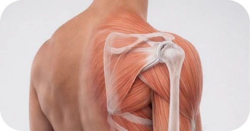
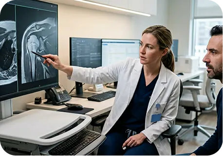
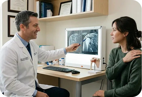
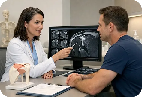
A SLAP tear is an injury to the cartilage ring (labrum) at the top of the shoulder socket, where the biceps tendon attaches. Your MRI or arthroscopic findings will classify the tear by type, severity, and whether the biceps anchor is stable or detached. These details determine the most appropriate treatment path, whether that is arthroscopic SLAP repair, biceps tenodesis, or another approach tailored to your activity level and goals.

A. Type I â Fraying (Non-Surgical)
The superior labrum shows degenerative fraying along its free edge, but the labrum remains firmly attached to the bone, and the biceps anchor is intact. Type I tears are common incidental findings on MRI, particularly in patients over 40. They rarely require surgical intervention and are typically managed with physical therapy and activity modification.

The superior labrum and the biceps tendon anchor have detached from the underlying bone (glenoid). This creates an unstable biceps anchor, which can cause pain with overhead activity, deep shoulder aching, mechanical catching, and the characteristic âdead armâ sensation that throwing athletes describe. Type II tears are the most clinically significant SLAP lesion and the most common type requiring surgical treatment. They can be further classified by extension direction:

A displaced, bucket-handle tear of the superior labrum that can catch or lock in the joint. The biceps anchor itself typically remains intact. The torn flap is removed (debrided) during arthroscopy, and the biceps attachment is preserved.

Similar to a Type III tear, but the bucket-handle fragment extends into the biceps tendon itself. The degree of biceps tendon involvement determines whether repair, tenodesis, or partial debridement is most appropriate. When a significant portion of the biceps tendon is involved, biceps tenodesis may be preferred to avoid leaving a structurally compromised tendon in the joint.
The biceps anchor is where the long head of the biceps tendon attaches to the top of the shoulder socket, and its status drives every SLAP tear treatment decision. An intact anchor means the labrum is still connected to bone and may be manageable without surgery. A detached or unstable anchor (Type II) means that structural connection has been disrupted, which typically requires surgical stabilization to restore reliable shoulder function. Stability is assessed through MRI arthrogram, physical examination, and when needed, direct arthroscopic visualization.

Traumatic (Acute) SLAP Tear
A sudden injury to the superior labrum caused by a specific event: a fall onto an outstretched hand (FOOSH), a shoulder dislocation, a traction injury (such as catching a heavy object), or a direct blow to the shoulder. Traumatic SLAP tears are common in younger patients and often involve a clean detachment of the biceps anchor (Type II), which tends to have a favorable prognosis when repaired promptly.
Â
Repetitive Overhead / Throwing Mechanism
In overhead athletes â particularly baseball pitchers, softball players, swimmers, volleyball players, and tennis players â SLAP tears develop through repetitive microtrauma. The âpeel-backâ mechanism occurs during the late cocking phase of the throwing motion, where the biceps tendon peels the superior labrum away from the glenoid. Internal impingement (also called posterior internal impingement) occurs when the rotator cuff compresses against the posterosuperior labrum during the acceleration and follow-through phases. These repetitive forces gradually weaken the biceps anchor until it detaches.
Â
Degenerative Change
In patients over 40, the superior labrum can develop age-related fraying and weakening (Type I changes). When a Type I labrum is then subjected to a minor injury or repetitive stress, it may progress to a Type II detachment. Degenerative SLAP changes are treated differently from traumatic tears in younger patients â biceps tenodesis is often preferred over repair in this population because the tissue quality may not support reliable anchor healing.
Standard MRI can miss SLAP tears because the superior labrum is a small structure and distinguishing a true tear from normal variants requires high contrast resolution.Â
Â
An MRI arthrogram, where contrast dye is injected into the joint before imaging, significantly improves diagnostic accuracy by filling the joint space and highlighting labral detachment that standard MRI might miss.Â
Â
If your standard MRI is inconclusive but symptoms strongly suggest a SLAP tear, your surgeon may recommend an MRI arthrogram or CT arthrogram for definitive evaluation.

SLAP tears frequently occur alongside other shoulder conditions. Your MRI or arthroscopic evaluation may reveal:
Â
Â
When multiple structures are compromised, all affected pathology is typically addressed during a single arthroscopic procedure to avoid the need for a second surgery.

*Same-day consultations may be available

MRI arthrogram provides valuable structural information but does not establish a complete diagnosis on its own. A SLAP tear must be interpreted alongside symptoms, physical examination findings, and functional demands. Specialized tests such as the O’Brien test, biceps load test, and dynamic labral shear help correlate imaging with clinical presentation. In some cases, the definitive diagnosis is made during diagnostic arthroscopy, where the surgeon can directly visualize the labrum and assess biceps anchor stability under conditions that MRI cannot replicate.

The superior labrum is pulled on by the biceps tendon with most every arm motion, which means a Type II tear with labral detachment is unlikely to reattach and heal on its own. Physical therapy can reduce pain and improve surrounding muscle strength, allowing some patients to function at an acceptable level, but it cannot reattach the labrum to bone or restore a stable biceps anchor. A comprehensive evaluation by a shoulder specialist will determine whether your tear can be managed conservatively or requires structural repair.

Not all SLAP tears progress at the same rate. Type I (fraying) tears may remain stable for years. However, Type II tears with an unstable biceps anchor carry specific risks when left untreated:
Â

When a SLAP tear progresses, the functional consequences depend on your demands:
For throwing and overhead athletes:
Â
Â
For working adults:
Â
Â
For active adults and recreational athletes:
Â
Â
Addressing a SLAP tear before compensatory damage develops to the rotator cuff or biceps tendon generally leads to more predictable surgical outcomes and a faster return to full activity.
Appropriate treatment for a SLAP tear depends on multiple factors: the tear type and severity, biceps anchor stability, how the tear developed, your age and activity demands, and your short- and long-term shoulder goals. The treatment decision is particularly nuanced for SLAP tears because the choice between repair, tenodesis, and debridement depends heavily on who you are and what you need your shoulder to do.
For some patients â particularly those with Type I (fraying) tears or low-demand lifestyles â structured physical therapy and activity modification can reduce pain and improve shoulder function without surgery. It is important, however, to understand what therapy can and cannot accomplish for a SLAP tear.
A well-designed rehabilitation program can:
Â
Â
Many patients experience meaningful improvement within 6â12 weeks when therapy is structured and progressive.


Physical therapy alone may not be appropriate when:
Â
Â
For overhead athletes and active working adults, the decision to move beyond therapy is often practical: the shoulder is no longer performing at the level your sport, training, or career demands. If strength and function have not returned after a structured rehabilitation program, or if injections provide shorter relief each time, this reflects the limits of non-surgical care and may indicate that structural repair or tenodesis is needed. A comprehensive evaluation will determine whether conservative management remains appropriate for your specific tear and what approach is most likely to restore reliable shoulder function.

When a Type II SLAP tear causes pain, mechanical symptoms, or compromises shoulder function â and conservative care has not restored reliable performance â arthroscopic SLAP repair may be recommended to reattach the detached labrum and biceps anchor to the glenoid bone.
Arthroscopic SLAP repair is a surgical procedure that reattaches the superior labrum â the cartilage ring at the top of the shoulder socket â back to the underlying bone. The surgery restores the biceps tendon anchor, which is the point where the long head of the biceps tendon originates from the top of the glenoid.
Â
The procedure is performed through small incisions (typically 3â4 portals) using a camera and specialized instruments, allowing the surgeon to directly visualize the labrum, assess the biceps anchor, and perform the repair with minimal tissue disruption.

SLAP repair is generally most appropriate for:
Â
Â
The decision between SLAP repair and biceps tenodesis is individualized. Your surgeon considers your age, activity demands, tissue quality, and the specific characteristics of your tear to recommend the approach most likely to produce a durable result.
SLAP repair is performed arthroscopically under regional anesthesia with sedation or general anesthesia.
Â
During surgery, the surgeon:
A. Evaluates the Labrum and Biceps Anchor
The arthroscope allows direct assessment of the superior labrum, biceps anchor stability, and any associated pathology (rotator cuff tears, Bankart lesions, cartilage damage). The tear type (IIA, IIB, IIC, III, IV) is confirmed, and the surgical plan is finalized based on what is found.
Â
B. Prepares the Labrum and Glenoid
The detached labrum is mobilized and the underlying glenoid bone is debrided to a fresh, bleeding surface. This bone preparation is essential â it creates the biological environment needed for the labrum to heal back to bone.
Â
C.Reattaches the Labrum with Suture Anchors
Small biocomposite or bioabsorbable suture anchors are placed into the glenoid bone at the superior rim. Sutures from these anchors are passed through the labral tissue and tied to secure the labrum back to its anatomic position. The number and placement of anchors depends on the size and extension of the tear â typically 1â3 anchors for a standard Type II repair.
Â
D. Confirms Repair Stability
The repair is inspected under direct visualization. The surgeon tests the biceps anchor to confirm that the labrum is securely reattached and that the biceps tendon origin is stable.
Â
The procedure typically takes approximately 60â90 minutes, depending on complexity and whether combined procedures are performed. Incisions are typically only a few millimeters in size.

When the superior labrum heals back to the glenoid:
Â
Â
For throwing athletes, successful SLAP repair preserves the native biceps anchor, which is critical for the deceleration phase of the throwing motion. This is the primary reason repair is often preferred over tenodesis for young competitive throwers.

Biceps tenodesis is an alternative surgical approach for SLAP tears that addresses the pain generator â the damaged biceps anchor â by detaching the long head of the biceps tendon from its origin at the superior labrum and reattaching it to a new position on the humerus (upper arm bone). Rather than repairing the labrum back to the glenoid, tenodesis eliminates the intra-articular source of pain while preserving biceps muscle strength and arm cosmesis.
Biceps tenodesis is generally considered for:
Â
Biceps tenodesis can be performed arthroscopically, through a small open incision, or as a combination. The biceps tendon is released from its attachment at the superior labrum, and the damaged or degenerative labral tissue is debrided. The tendon is then secured to the humerus â either in the bicipital groove or at the subpectoral level â using a fixation device (typically an interference screw, suture anchor, or cortical button).
Â
The torn or frayed superior labral tissue is smoothed (debrided) to eliminate any remaining mechanical symptoms. The procedure typically takes 45â75 minutes.

Tenodesis eliminates the intra-articular biceps anchor â which is the primary source of pain in most SLAP tears â while preserving biceps muscle strength and normal arm appearance. Because the tendon is secured to a robust fixation point on the humerus rather than to a small rim of labral tissue, tenodesis fixation is generally strong and reliable.
Â
Published data show high satisfaction rates and reliable pain relief with biceps tenodesis for SLAP tears, particularly in patients over 35.

Recovery after biceps tenodesis generally follows a similar timeline to SLAP repair, with some differences:
Â

One of the most common and important questions for patients diagnosed with a Type II SLAP tear is whether to pursue arthroscopic SLAP repair or biceps tenodesis. This decision is not one-size-fits-all â the right choice depends on your age, activity demands, tissue quality, and goals.
Age and Tissue Quality
Younger patients (typically under 35â40) with good labral tissue quality are generally better candidates for SLAP repair. The labrum in younger patients tends to heal more reliably to bone. As patients move into their 40s and beyond, degenerative changes in the labrum can reduce the healing potential of a repair, making tenodesis a more predictable option.
Â
Activity Demands and Sport
For overhead throwing athletes, the native biceps anchor plays a role in the deceleration phase of the throwing motion, making SLAP repair the anatomy-preserving option. However, whether repair or tenodesis produces better return-to-throwing outcomes remains actively debated, and the decision is made collaboratively between the athlete, surgeon, and sports medicine team. For non-throwing overhead athletes such as swimmers, volleyball players, and CrossFit athletes, both repair and tenodesis can produce excellent outcomes, with tenodesis offering a more predictable recovery and lower retear risk. For non-overhead workers and recreational athletes, tenodesis often provides reliable pain relief with a straightforward recovery.
Â
Failure Rates
Published literature shows that SLAP repair has a higher failure rate than biceps tenodesis, particularly in patients over 35â40 and in cases with degenerative tissue quality. Failure after SLAP repair is typically managed with conversion to tenodesis, which produces good outcomes as a revision procedure.
Â
Strength and Cosmesis
Both procedures preserve biceps muscle strength. Tenodesis maintains normal arm appearance (biceps contour). Tenotomy â simply cutting the biceps tendon without reattaching it â may result in a visible change in biceps contour (sometimes called a âPopeye deformityâ), which is why tenodesis is generally preferred over tenotomy for active and cosmetically-conscious patients.
Â
At The Joint Preservation Center, both procedures are performed regularly, and the recommendation is based on what is most likely to produce a durable, functional result for your specific situation â not a one-size-fits-all protocol.
For Type I SLAP tears (labral fraying without detachment) and Type III SLAP tears (bucket-handle tears with an intact biceps anchor), debridement â the removal or smoothing of damaged labral tissue â may be the most appropriate surgical treatment. Debridement removes the mechanical source of catching or pain without disrupting the biceps anchor.
Â
Recovery after labral debridement is typically faster than repair or tenodesis. Most patients begin active motion within 1â2 weeks and return to full activity within 6â10 weeks.
Biceps tenotomy involves simply cutting (releasing) the long head of the biceps tendon from its attachment at the superior labrum, without reattaching it to a new position. The released tendon retracts into the arm.
Â
Tenotomy is a simpler and faster procedure than tenodesis, and may be considered for lower-demand patients where cosmetic appearance (biceps contour) is not a primary concern. The main trade-off is a potential visible change in biceps muscle shape (âPopeye deformityâ). For active patients who value arm appearance and strength, tenodesis is generally preferred.
Not every SLAP tear can be addressed with a straightforward arthroscopic repair or tenodesis alone. Combined pathology, complex tear patterns, bilateral involvement, and failed prior repairs each present distinct challenges that require advanced techniques and specialized experience.
Â
At The Joint Preservation Center, we evaluate the full spectrum of labral pathology and offer the appropriate procedure for your specific situation, rather than a one-size-fits-all approach.
SLAP Repair with Rotator Cuff Repair
Overhead athletes and patients with traumatic injuries frequently present with both a SLAP tear and a rotator cuff tear. When both structures are compromised, addressing only one may leave residual pain or instability. We evaluate each case to determine whether the SLAP component requires repair, tenodesis, or debridement, and perform the combined procedure to restore both labral stability and cuff integrity in a single operation.
Â
SLAP Repair with Bankart Repair
When the shoulder has dislocated, the labrum may tear at both the top (SLAP) and front (Bankart) of the socket. Combined SLAP and Bankart repair addresses the full circumference of labral damage to restore shoulder stability. This is particularly common in young patients with traumatic dislocations.
Â
SLAP Debridement with Biceps Tenodesis
For patients with a SLAP tear combined with significant biceps tendon pathology (partial tearing, tendinosis, or subluxation), the damaged labral tissue is debrided and the biceps is relocated via tenodesis. This combined approach eliminates both the labral and biceps pain generators in a single procedure.

A SLAP repair that does not heal â whether due to a retear, anchor failure, or persistent symptoms â can leave patients with ongoing pain, mechanical symptoms, or inability to return to sport or work. For overhead athletes and active working adults, a failed SLAP repair is especially frustrating because the recovery investment has already been made.
Â
When Is Revision Surgery Appropriate?
Revision SLAP surgery may be considered when imaging or clinical examination confirms repair failure or retear, symptoms have not resolved after appropriate postoperative rehabilitation, and the clinical picture suggests that a second procedure has a reasonable chance of improving function. We evaluate revision cases with advanced imaging and a thorough assessment of what may have contributed to the initial failure â including tissue quality, anchor placement, rehabilitation adherence, and patient biology.
Â
Signs of SLAP Repair Failure to Watch For
Â
Revision Approaches
Depending on the revision scenario, we may:
Â
The revision plan is determined by what the labral tissue, biceps tendon, and joint can support at the time of reevaluation.

Some patients â particularly overhead athletes and workers with heavy bilateral arm demands â present with SLAP tears in both shoulders. Bilateral surgery is typically staged: one shoulder is addressed first, and the second is treated after the first has recovered enough for the patient to manage daily activities. We develop a surgical and rehabilitation plan that sequences bilateral procedures to minimize cumulative downtime.

Recovery timelines after SLAP surgery differ significantly between arthroscopic SLAP repair and biceps tenodesis â and are substantially affected by whether concurrent procedures (rotator cuff repair, Bankart repair, decompression) were performed at the same time. For competitive pitchers, the interval throwing program timeline governs return to sport, and this is the longest component of SLAP repair recovery.
Most patients experience moderate discomfort in the first several days following SLAP repair or biceps tenodesis, which is managed with a combination of regional anesthesia (nerve block), oral medication, and icing. A sling is worn to protect the repair.
Â
Â
Pain typically improves significantly within the first 1â2 weeks. Sleeping in a slightly upright position (recliner or wedge pillow) is often most comfortable during the early phase.

Phase 1: Protection and Passive Motion (Weeks 0â4/6)
Gentle, guided movement begins early to prevent stiffness while protecting the repair. The repaired labrum is not yet healed to bone.
Â
Phase 2: Active Motion (Weeks 4/6â8/10)
You begin moving the shoulder under your own power as healing progresses. Active range of motion is gradually restored.
Â
Phase 3: Strengthening (After ~10â12 Weeks)
Controlled strengthening exercises begin once the labrum has biologically started to integrate into the bone. Rotator cuff and scapular strengthening form the foundation.
Â
Phase 4: Sport-Specific and Throwing Rehabilitation (After ~12â16 Weeks)
For overhead athletes and throwers, sport-specific rehabilitation begins once baseline strength milestones are met. For pitchers and throwing athletes, this phase includes an interval throwing program (ITP) â a structured, progressive throwing protocol that gradually increases distance, effort, and volume over several months.

Drive?
Usually when you are out of the sling and off narcotic pain medication â typically 3â6 weeks after surgery.
Â
Return to desk work?
Often within 1â2 weeks, depending on comfort.
Â
Return to manual labor?
Typically 3â5 months, depending on overhead demands.
Â
Return to non-throwing sports (swimming, CrossFit, tennis, volleyball)?
Gradual return typically begins around 4â6 months, with full participation at 6â9 months.
Â
Return to throwing (baseball, softball, football)?
An interval throwing program typically begins around 4â5 months. Competitive throwing return ranges from 9â12 months depending on sport, competition level, and healing.
Â
Timelines vary based on tear type, procedure (repair vs. tenodesis), tissue quality, and individual healing response.
Desk work and computer-based roles:
Most patients return to desk work within 1â2 weeks, working one-handed or with limited arm use initially. Remote work may allow an even earlier transition.
Â
Supervisory, managerial, or client-facing roles:
If your work does not require physical lifting, return within 1â2 weeks is typical. Driving is usually possible once the sling is discontinued.
Â
Manual labor, construction, trades, or physical occupations:
Return to light duty may be possible within 2â3 months. Return to full overhead or heavy lifting duties typically requires 4â6 months depending on healing and whether repair or tenodesis was performed.
Â
Military service members:
Return to full duty typically requires 6â9 months. We provide documentation for medical boards, fitness assessments, and return-to-duty clearance as needed.
Â
Work restrictions documentation:
We provide work restriction letters and return-to-work clearance documentation as needed for your employer or occupational health department.
These timelines are approximations. Your specific return-to-sport plan is developed collaboratively between your surgeon and your physical therapist based on objective strength and functional milestones.
Preparing your home and support system before surgery helps the early recovery period go more smoothly. Because you will be in a sling and restricted from using your operative arm for several weeks, advance planning makes a meaningful difference:
Â
Â
Patients who plan ahead consistently report a smoother, less stressful early recovery.
Week 1:
Sling wear at all times. Pain management with medication, icing, and rest. Begin gentle pendulum exercises as directed. Most discomfort resolves significantly by day 5â7.
Â
Weeks 2â3:
First postoperative visit. Passive range-of-motion exercises with a therapist begin. Pain continues to improve. Desk work may resume if comfortable.
Â
Weeks 4â6:
Gradual progression of passive and assisted motion. Sling is typically discontinued around week 4â6 depending on procedure and repair security. Driving may resume once the sling is off and narcotic medication is no longer needed.
Â
Weeks 6â10:
Transition from passive to active motion. The shoulder begins moving under its own power. Daily activities become noticeably easier.
Â
Weeks 10â16:
Strengthening begins. Controlled resistance exercises rebuild rotator cuff and scapular strength. Light recreational activity may resume. Throwing athletes may begin a light toss program at the end of this phase.
Â
Months 4â6:
Progressive strengthening and sport-specific rehabilitation. Interval throwing program progresses for throwing athletes. Many non-throwing patients return to most activities during this window.
Â
Months 6â12:
Continued strength gains. Throwing athletes progress to competitive throwing. Full recovery and confidence with demanding activities. Strength and performance can continue improving for up to a year after surgery.

Modern arthroscopic repair techniques are associated with:
High rates of pain improvement
Significant gains in strength
Improved functional outcome scores
High patient satisfaction
1
We evaluate your symptoms, shoulder strength and movement, review any imaging (including MRI arthrogram findings), and consider your goals, activity demands, and sport. If advanced imaging is needed, we can order an MRI arthrogram for definitive evaluation.
2
We explain whether rehabilitation, SLAP repair, biceps tenodesis, or another approach is most appropriate based on your tear type, age, activity level, and goals.
3
If surgery is recommended, our fellowship-trained shoulder surgeon performs the appropriate arthroscopic procedure â SLAP repair, biceps tenodesis, debridement, or combined approach â to address your specific pathology and restore shoulder function.
4
Our surgeons work closely with physical therapists to guide a structured rehabilitation program that restores motion and strength while protecting the repair. For throwing athletes, rehabilitation includes a sport-specific interval throwing program.
5
Rehabilitation progresses through milestones so you can confidently return to the activities that matter most â throwing, overhead sport, work, and daily life.
This long-term follow-up helps us understand how shoulders recover beyond the early healing period â including strength, function, return to sport, and return to throwing. These insights allow our surgeons to continually refine surgical planning and rehabilitation strategies to support durable shoulder performance over time.

1
Elite surgeons with decades of experience, incentivized to do the right thing
2
Prevent future surgeries
3
Heal with advanced, minimally invasive techniques
4
Preserve your natural joints, whenever possible
5
Seamless coordination from injury to recovery
6
Premium personalized care, made accessible
7
All patient outcomes tracked for 5 years
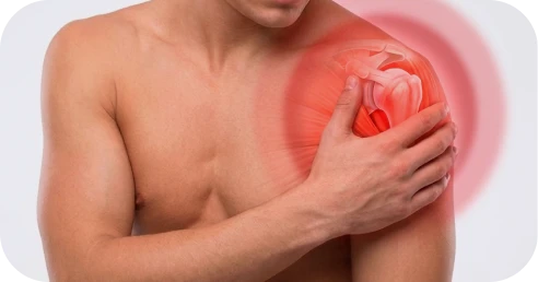
A SLAP tear â short for Superior Labrum Anterior to Posterior â is an injury to the cartilage ring (labrum) at the top of the shoulder socket. The superior labrum is where the long head of the biceps tendon attaches to the shoulder. When this area tears, it can cause pain, clicking, catching, weakness with overhead activity, and the characteristic âdead armâ sensation that many throwing athletes experience.
Â
SLAP tears are classified into four types (Type IâIV). Type II â where the labrum and biceps anchor detach from the underlying bone â is the most common type requiring surgical treatment.
SLAP tear symptoms vary depending on the tear type and severity. Common symptoms include:
Â
Â
Some SLAP tears develop gradually from repetitive overhead use. Others occur suddenly after a fall, traction injury, or shoulder dislocation.
SLAP tears are diagnosed by an orthopedic surgeon who specializes in shoulder conditions, often through a combination of:
Â
Â
Because imaging findings do not always perfectly match symptoms, the final diagnosis is based on a combination of physical examination, imaging, and how the shoulder is functioning.
SLAP tears are treated by orthopedic surgeons who specialize in shoulder and sports medicine conditions. If you are looking for the highest level of specialization, look for a fellowship-trained shoulder surgeon â someone who completed an additional year of advanced training focused specifically on complex shoulder conditions and procedures, including SLAP repair and biceps tenodesis.
Â
For throwing athletes, a surgeon with specific experience treating overhead and throwing athletes is especially important, as the repair-vs-tenodesis decision and rehabilitation approach may differ from the general population.
This is the most common question we hear from SLAP tear patients, and there is no single answer for everyone. The choice depends on your age, activity demands, tissue quality, and goals:
Â
Â
Both procedures produce good outcomes when matched to the right patient. Your surgeon will discuss the specific factors in your case and make a collaborative recommendation.
Â
For a detailed comparison, see the Repair vs. Tenodesis section above.
Outcomes after SLAP repair depend on tear type, patient age, tissue quality, and activity demands. Published data show:
Â
Â
At The Joint Preservation Center, we track outcomes for 5 years because the true success of SLAP tear surgery is not just early pain relief â it is whether the shoulder remains strong, functional, and reliable over time.
Recovery timelines depend on the procedure performed and your activity demands:
Â
Â
Full strength and confidence can continue improving for up to a year after surgery.
Patients do not feel pain during surgery. The procedure is typically performed under regional anesthesia with sedation or general anesthesia. Many patients also receive a regional nerve block, which keeps the shoulder numb for several hours after surgery.
Â
After surgery, moderate discomfort is expected during the first 48â72 hours and is managed with medication, icing, and sling support. Pain typically improves steadily over the first 1â2 weeks. Many patients find sleeping in a slightly upright position most comfortable during the early phase.
If a SLAP repair does not heal or symptoms return, the most common revision approach is conversion to biceps tenodesis. Tenodesis as a revision procedure generally produces good outcomes, with reliable pain relief and improved function. In some cases, a repeat repair may be possible if the tissue quality supports it.
Â
If you are experiencing persistent pain, clicking, dead arm, or functional limitations after a prior SLAP repair, a comprehensive evaluation can determine whether revision surgery is appropriate.
Return to throwing is one of the most important goals for overhead and throwing athletes after SLAP tear surgery. The timeline typically follows a structured interval throwing program (ITP) that begins around 4â5 months postoperatively and progresses gradually over several months.
Â
Return to competitive pitching typically ranges from 9â12 months. Return-to-throwing rates and velocity outcomes vary by age, competition level, tear type, and procedure. Your surgeon and physical therapist develop a throwing return plan based on objective strength and functional milestones, not arbitrary calendar dates.
Yes. SLAP tears frequently occur alongside other shoulder conditions, including rotator cuff tears, Bankart lesions (anterior labral tears from shoulder dislocation), Hill-Sachs lesions (bone impressions from dislocation), and biceps tendon damage. When multiple structures are compromised, all affected pathology is typically addressed during a single arthroscopic procedure.
Â
The Joint Preservation Center accepts most PPO insurance plans that have out-of-network benefits:
Â
If you have a PPO insurance plan with out-of-network benefits:
Â
Â
Note: If you have Medicare, Medicaid, TRICARE, or VA programs, or if your PPO does not have out-of-network benefits, you can still see our specialists and the surgery center will still be in-network. In this case, our specialists charge $250 for the initial office visit (all follow ups are included). Surgery is typically in the range of $6K â $8K depending on what you need done.
Â
If you are unsure whether your plan is accepted, our team can verify your coverage before your appointment.
*Same-day consultations may be available
*Online visits available