

*Online Consultations Available
*Online Consultations Available
Cartilage Repair | OATS |Â OCA | ACIÂ | MACI
OCD Fixation | Drilling | DeNovo | BioCartilage
TTO / Fulkerson | Trochleoplasty | MPFL | HTO
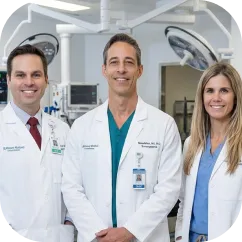
1,000+ Shoulder Surgeries performed
Rehab-IntegratedâĻ
Care
Outcomes Tracked for 5 Years
Articular cartilage â the smooth, white tissue that covers the ends of the bones in the knee joint â has no blood supply and virtually no capacity for self-repair. When cartilage is damaged, it does not grow back on its own. The type of damage you have determines which procedures are appropriate, which are not, and what the realistic outcomes are.
A focal chondral defect is an isolated area of cartilage loss on the femoral condyle, patella, or trochlear groove without involvement of the underlying bone. These defects are graded 1 through 4 by depth, with Grade 3 and Grade 4 lesions â partial to full-thickness cartilage loss, sometimes described as down to bone in a localized area â being the primary surgical candidates. Treatment options include microfracture, nanofracture, OATS, osteochondral allograft (OCA), ACI, MACI, BioCartilage, and DeNovo particulated juvenile cartilage.Â

An osteochondral defect involves both the cartilage surface and the underlying subchondral bone. Osteochondritis dissecans (OCD) is the most common cause, where a segment of bone beneath the cartilage loses its blood supply, becomes necrotic, and separates from the surrounding bone. OCD lesions are staged 1 through 4 by stability, with Stage 3 (partially detached) and Stage 4 (fully detached or loose body) being surgical indications.Â
Â
The defining surgical question is fixation vs. replacement: a viable, attached fragment can often be fixed in place with screws and bone grafting, while a detached or non-viable fragment requires osteochondral allograft (OCA) to restore the joint surface. Procedures include OCD fragment fixation with bioabsorbable screws, Herbert screws, or suture fixation; retrograde and antegrade drilling; bone grafting; and OCA.

Patellofemoral cartilage defects affect the joint between the kneecap and the trochlear groove, and frequently occur alongside patellofemoral malalignment â an abnormal tracking pattern that concentrates stress on specific cartilage areas. Cartilage repair without addressing the underlying malalignment frequently fails, which is why tibial tubercle osteotomy (TTO/Fulkerson/AMZ) is often performed concurrently with cartilage restoration in this population. Procedures include ACI, MACI, OATS, and OCA for the patella and trochlea; TTO for malalignment correction; MPFL reconstruction for instability-driven cartilage damage; and trochleoplasty for trochlear dysplasia with cartilage involvement.

Some patients have cartilage damage that has been evaluated by another surgeon and told they need knee replacement â but they are in their 30s, 40s, or 50s, are highly active, and are not ready or willing to undergo replacement surgery. For these patients, cartilage restoration procedures represent a genuine alternative that can restore joint function, delay or prevent knee replacement, and preserve the native knee for years or decades.
Â
Joint preservation is not appropriate for every patient â diffuse, end-stage arthritis throughout the knee cannot be addressed with focal cartilage procedures. But for patients with focal or osteochondral defects in an otherwise preserved joint, joint preservation surgery at The Joint Preservation Center provides a meaningful and evidence-supported pathway to avoid or significantly delay replacement.

*Same-day consultations may be available

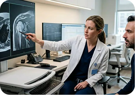
Even though an MRI provides critical structural information about cartilage damage grade, defect size, OCD staging, and bone involvement, it does not establish a complete diagnosis on its own. The appropriate treatment depends on multiple factors beyond imaging â including symptoms, physical examination, activity level, age, limb alignment, and concurrent ligament or meniscal pathology. Two patients with identical MRI findings can require completely different procedures. A specialist evaluation that integrates all clinical dimensions, alongside specialized imaging when indicated, is required to determine the right surgical approach.Â

Articular cartilage is graded on the International Cartilage Repair Society (ICRS) scale and the Outerbridge classification, which are closely related:

A. Grade 1
Cartilage softening and surface irregularity. The cartilage surface is intact but mechanically compromised. Almost always managed conservatively.


Full-thickness cartilage loss exposing the subchondral bone. The cartilage is gone down to bone â sometimes described as ‘bone on bone’ in a localized area. Grade 4 defects are the primary surgical target for cartilage restoration procedures.

A. Medial femoral condyle (MFC)
The most common location for focal chondral defects and OCD lesions. The weight-bearing surface of the inner femoral condyle is under high compressive load with every step. MFC defects are the most commonly treated with OATS, OCA, ACI, and MACI.

B. Lateral femoral condyle (LFC)
The outer condyle. Lateral OCD lesions are common in younger athletes. Similar procedure options to MFC.

C. Patella (kneecap)
The undersurface of the kneecap bears high contact pressure with knee flexion. Patellar cartilage defects often occur with patellar malalignment or instability. ACI, MACI, and OCA are used; TTO is frequently combined.

D. Trochlear groove
The groove in which the kneecap tracks. Trochlear cartilage defects are less commonly recognized but equally symptomatic, with pain concentrated at the front of the knee with activity. Procedures: MACI, OCA; trochleoplasty when trochlear dysplasia coexists.
Size is one of the most important factors in procedure selection for focal chondral defects.
Â
When MRI shows not just cartilage damage but bone involvement beneath the cartilage â subchondral bone edema, cystic changes, or frank osteochondral defect â the management changes.
Â
For OCD lesions specifically, MRI staging determines whether fixation or replacement is the appropriate surgical approach.
Â

For patients with patellar or trochlear cartilage defects, the TT-TG (tibial tubercle to trochlear groove) distance measured on CT scan quantifies how far off-center the kneecap tracks relative to the groove. A TT-TG distance greater than 15â20mm indicates significant malalignment â and when cartilage restoration is performed without correcting the malalignment, the mechanical overload on the repaired cartilage leads to failure.
When TT-TG is elevated, tibial tubercle osteotomy (TTO) â in which the tibial tubercle attachment of the patellar tendon is surgically moved to correct the tracking vector â is performed concurrently with or before cartilage restoration.
No â not in any clinically meaningful sense. Articular cartilage has no blood supply and no meaningful capacity for self-repair. Full-thickness defects (Grade 4) and unstable OCD lesions do not regenerate on their own. What the body can produce is fibrocartilage â a scar-like tissue that is mechanically inferior to native hyaline cartilage and typically breaks down over time under compressive load. This is one of the reasons microfracture, which relies on fibrocartilage fill, has documented durability limitations for large defects and high-demand patients.

For athletes, active adults, and working-age patients, untreated cartilage damage produces progressive loss of sport and high-impact capacity, increasing pain with stairs and daily activity, and eventual loss of the joint preservation window â the period during which restoration is still possible before diffuse arthritis develops. Timing matters: a focal Grade 4 defect treated in an otherwise well-preserved joint produces better outcomes than the same defect treated after years of secondary damage have accumulated. Earlier evaluation expands options.
Conservative management â cortisone injections, hyaluronic acid (gel) injections, PRP, physical therapy, activity modification, unloading bracing, and NSAIDs â addresses the inflammatory and symptomatic consequences of cartilage damage. It does not restore the cartilage surface, fill the defect, or stabilize an OCD fragment.
Â
For patients who have completed a defined course of conservative management without achieving adequate functional improvement, the signals that surgical evaluation is appropriate are specific:
Â
Knee cartilage restoration encompasses a spectrum of procedures matched to defect size, depth, location, OCD staging, and patient factors including age, activity level, and the status of the surrounding joint. At The Joint Preservation Center, we evaluate every patient comprehensively before recommending a procedure â matching the biology of the repair to the biology of the defect.
Microfracture is the most commonly performed cartilage procedure â arthroscopic creation of small perforations in the subchondral bone plate beneath the cartilage defect to stimulate bleeding and marrow-derived stem cell recruitment, producing a fibrocartilage repair fill.

Microfracture produces fibrocartilage, not hyaline cartilage â mechanically inferior and less durable under long-term loading. Published evidence shows good short-term results but higher deterioration rates over time, particularly for large defects, high-demand athletes, and patellofemoral lesions. For patients who have undergone microfracture and continue to have symptoms, revision with OATS, OCA, ACI, or MACI is the appropriate next step. Nanofracture refines the technique with smaller, more precisely placed perforations that better preserve the subchondral bone plate. BioCartilage augmented microfracture adds a particulated cartilage matrix scaffold to improve the quality of the fibrocartilage fill. Both are available as first-line or augmentation options for smaller defects.
OATS (osteochondral autograft transplantation system) â sometimes called mosaicplasty when multiple plugs are used â transfers cylindrical osteochondral plugs (bone and overlying hyaline cartilage) from a non-weight-bearing area of the patient’s own knee to fill the cartilage defect. Because autograft uses the patient’s own tissue, the transplanted cartilage is hyaline cartilage â mechanically superior to fibrocartilage.

The plug harvest site (typically the periphery of the lateral femoral condyle or the intercondylar notch) represents a donor site â limited in the total number of plugs that can be safely harvested. For defects larger than 4 cm squared, donor site constraints make OATS insufficient, and OCA becomes the preferred option. Donor site pain or discomfort is reported in some patients.
Fresh osteochondral allograft (OCA) transplantation uses a matched, fresh donor graft â supplied by a tissue bank â to replace large cartilage and bone defects with a composite osteochondral unit that is anatomically matched to the patient’s joint geometry. Unlike OATS, OCA can address large defects without donor site limitations.

Fresh osteochondral allograft requires tissue bank matching â the donor graft is matched to the patient’s knee size and the geometry of the defect from fresh cadaveric tissue. The fresh (not frozen) state preserves viable chondrocytes that integrate more reliably than processed allograft. Tissue availability may affect surgical timing, and patients are counseled about the scheduling implications at consultation.
Autologous chondrocyte implantation (ACI) and its matrix-induced evolution (MACI) use the patient’s own cartilage cells â harvested arthroscopically in a first-stage procedure, expanded in a laboratory, and then implanted at the defect site in a second-stage open procedure â to regenerate hyaline-like cartilage at the defect.
Traditional ACI injects expanded chondrocytes beneath a periosteal or collagen membrane cover. MACI (matrix-induced ACI, the FDA-approved commercial product) seeds the expanded chondrocytes onto a collagen scaffold membrane that is then sized and implanted as a single construct. MACI is technically simpler to implant than traditional ACI and produces equivalent or superior clinical results in most published comparisons.

ACI and MACI require two surgical procedures: Stage 1 is a brief arthroscopic cartilage biopsy harvest (outpatient, 30 minutes). The harvested cells are sent to a laboratory where they are expanded over 4â6 weeks. Stage 2 is the implantation procedure (outpatient, 60â90 minutes for isolated defects). Recovery after Stage 2 is the longer rehabilitation period. The two-stage commitment is a meaningful consideration that is discussed explicitly at consultation.
DeNovo NT is an off-the-shelf particulated cartilage graft derived from donor juvenile cartilage. Juvenile chondrocytes have higher proliferative and repair capacity than adult chondrocytes, and the particulated format eliminates the need for cell expansion and a second surgical stage. DeNovo is most appropriate for small to medium defects, typically less than 2.5 cm squared, where the graft can be retained with fibrin glue and a membrane cover. It provides a single-stage alternative to ACI/MACI for selected cases, including patellar and trochlear defects, and is well-established in the cartilage restoration literature.
When an OCD fragment is still biologically viable â intact, partially detached, with good bone quality and no cystic degeneration at the bed â surgical fixation preserves the patient’s own native cartilage rather than replacing it with a donor graft. This is the preferred approach when the anatomy supports it.

For patients with patellofemoral cartilage defects combined with patellar malalignment (elevated TT-TG distance, patella alta, or patellar tilt), cartilage restoration alone is inadequate â the mechanical cause of the cartilage damage must be corrected simultaneously. Tibial tubercle osteotomy achieves this by moving the tibial tubercle (the attachment point of the patellar tendon) to correct the kneecap’s tracking vector.
Â
TTO is almost always performed combined with cartilage restoration (ACI, MACI, or OCA) at the same surgical session. Performing TTO alone without addressing the cartilage defect leaves the structural cartilage damage unresolved.

Trochlear dysplasia is an abnormal shallowness or flatness of the trochlear groove â the groove in which the kneecap tracks. When trochlear dysplasia coexists with trochlear cartilage damage, trochleoplasty (surgical deepening of the groove to normal depth) addresses the anatomic cause of both the instability and the abnormal contact patterns that damage the trochlear cartilage. Trochleoplasty is the most technically demanding procedure in the patellofemoral reconstruction spectrum and requires specialized surgical experience.
When patellar instability (dislocation or subluxation) has produced cartilage damage on the medial patellar facet or the lateral trochlea, MPFL (medial patellofemoral ligament) reconstruction addresses the instability while cartilage restoration (ACI, MACI, OCA) addresses the cartilage damage â both performed in the same or staged surgical procedure.
For patients with varus (bow-legged) alignment and medial compartment cartilage defects, the mechanical overload on the medial femoral condyle cannot be resolved by cartilage restoration alone. High tibial osteotomy (HTO) corrects the varus alignment by shifting the body’s weight-bearing axis away from the damaged medial compartment, reducing the mechanical load on the cartilage restoration. HTO is frequently combined with cartilage restoration procedures in patients with malalignment and medial focal defects.
For patients with OCD lesions, the most important surgical decision is whether the OCD fragment can be fixed in place (preserving the patient’s own native cartilage) or whether it must be replaced with an osteochondral allograft. This decision depends on fragment viability, size, and the condition of the bone bed.
Â
At The Joint Preservation Center, OCD fixation decisions are made using MRI with dedicated OCD sequences, CT scan for bone bed assessment and cyst characterization, and intraoperative arthroscopic assessment of fragment viability â including probing of the fragment and evaluation of the bone bed under direct visualization.
Osteochondritis dissecans (OCD) is one of the most common causes of knee pain in active adolescents â particularly in young athletes in high-impact and pivoting sports including soccer, basketball, gymnastics, hockey, and wrestling. In skeletally immature patients (those with open growth plates), OCD has a higher capacity for biological healing with conservative management than in adults â but when conservative management fails, surgical treatment requires specific expertise in pediatric and adolescent knee reconstruction.
Stable OCD lesions (Stage 1 and early Stage 2) in skeletally immature patients are typically managed conservatively first: activity restriction, non-weight-bearing or limited weight-bearing, and serial MRI monitoring at 3â6 month intervals to assess healing. The open growth plates in younger patients provide a biological environment more favorable for OCD healing than in adults â and surgery is appropriately deferred until conservative management has been given a reasonable chance.
Â
Conservative management is considered to have failed when: follow-up MRI at 3â6 months shows no healing or progression, the patient develops mechanical symptoms (catching, locking) indicating fragment instability, or a Stage 3 or 4 lesion is identified at initial presentation.
In skeletally immature patients with open growth plates, surgical technique must be adapted to avoid growth plate damage. Retrograde drilling â approaching the OCD bed from outside the joint without crossing the growth plate â is the technique of choice for stimulating healing while protecting the physis. When fragments require fixation, screw placement is planned to avoid the growth plates.
Juvenile OCD recovery â whether non-operative or surgical â typically requires 3â6 months before return to sport. For young athletes, surgery timing relative to the competitive season is an important planning consideration: fall sport athletes benefit from spring or early summer surgery; winter sport athletes benefit from late spring or summer surgery. School attendance is typically possible within 1â2 weeks of surgery, with PE restriction for the duration of the protected recovery.
The most important principle in knee cartilage procedure selection is matching the procedure to the defect â defect size, location, depth, bone involvement, OCD staging, patient age, and activity demands all determine which procedure produces the best outcomes for an individual patient. There is no universally superior cartilage procedure; there is only the appropriate procedure for the specific clinical situation.
Microfracture produces fibrocartilage fill that provides good short-term results for small defects in younger patients, but has documented durability limitations at medium and long-term follow-up â particularly for large defects and high-demand athletes. OATS provides hyaline cartilage transfer with bone-to-bone healing, producing more durable results for small to medium defects. OATS is generally preferred over microfracture when the defect size and location support plug transfer.
OATS uses the patient’s own tissue, avoiding donor-related concerns, but is limited by donor site availability to small-medium defects. OCA has no donor site constraints and can address large defects with anatomically matched composite grafts. For defects larger than 4 cm squared, OCA is the preferred reconstruction. OCA requires tissue matching and may have scheduling implications due to tissue bank logistics.
Traditional ACI and MACI both use the patient’s own expanded chondrocytes. MACI uses a collagen scaffold delivery vehicle, is technically simpler to implant, and has equivalent or superior outcomes in most published comparisons. MACI is the commercially available FDA-approved product. Both require a two-stage procedure. ACI and MACI are particularly well-validated for patellofemoral cartilage defects where OCA and OATS are more technically challenging.
MACI provides a cell-based hyaline-like cartilage repair using the patient’s own cells â no donor tissue concerns, but two-stage surgery and extended recovery. OCA provides an immediate single-stage reconstruction with proven hyaline cartilage, but requires tissue matching and donor tissue. For larger defects, OCA and MACI are the primary competitors â the choice depends on defect size, patient preference for autologous vs. allograft tissue, and the specific anatomic geometry.
BioCartilage (particulated extracellular cartilage matrix augmenting the microfracture site) and nanofracture (refined micro-perforation technique) both represent improvements over standard microfracture for small defects. The evidence base for these refinements continues to accumulate. For small defects where microfracture is the size-appropriate choice, BioCartilage augmentation is available as an enhancement to standard technique.
Fragment fixation preserves the patient’s own native cartilage and bone when the fragment is viable. OCA replaces the OCD fragment with a matched donor graft when the native fragment is non-viable, detached, or too large for stable fixation. Fragment fixation is always preferred when biologically feasible â preserving native tissue is preferable to transplanting donor tissue. The decision is made based on fragment viability assessment. See Section 9 for the complete OCD fixation vs. allograft decision framework.
The CARTIHEAL AGILI-C (Agili-C) is an FDA-approved, off-the-shelf aragonite-based scaffold for the treatment of chondral and osteochondral knee defects â including patients with mild to moderate osteoarthritis (Kellgren-Lawrence grade 0-3) who are not yet candidates for knee replacement.Â
Outcome tracking after cartilage restoration surgery goes beyond pain relief. We measure cartilage-specific results using validated instruments:Â Â
Â
Â
We track these outcomes systematically for 5 years after surgery. This allows us to understand individual healing trajectories and to continually refine patient selection and surgical technique.
Isolated focal chondral defects in an otherwise intact knee are the most straightforward cartilage cases. More commonly, cartilage damage occurs alongside other structural problems that must be assessed and addressed as part of the surgical plan.
The medial meniscus and the medial femoral condyle cartilage are functionally linked â meniscal loss accelerates medial condyle cartilage deterioration by eliminating the shock absorption and stress distribution that the meniscus provides. When a meniscal tear is identified alongside a cartilage defect, meniscal preservation through repair (rather than partial meniscectomy) is the highest priority â protecting the cartilage restoration from the overload that would follow meniscal removal.
ACL instability generates abnormal joint mechanics with every pivot and giving-way episode, progressively damaging articular cartilage. Cartilage damage after ACL injury is common. When both an ACL tear and a cartilage defect are present, ACL reconstruction and cartilage restoration can often be performed at the same surgical session â addressing both structural problems simultaneously.

Varus (bow-legged) alignment concentrates weight-bearing forces on the medial compartment, mechanically overloading medial femoral condyle cartilage. Cartilage restoration without addressing the alignment overload has a high failure rate. High tibial osteotomy (HTO) corrects the varus alignment â shifting load away from the cartilage restoration â and is performed concurrently or as a first stage before cartilage restoration in patients with significant varus malalignment.

Patellar instability â subluxation or dislocation of the kneecap â produces characteristic cartilage damage on the medial patellar facet and the lateral trochlear wall. MPFL reconstruction addresses the instability mechanism while cartilage restoration addresses the damaged surface. In patients with elevated TT-TG distance, tibial tubercle osteotomy (TTO) corrects the malalignment at the same time.

Knee replacement (TKA) is an effective treatment for diffuse, end-stage knee arthritis â but it is not appropriate for patients with focal cartilage defects in an otherwise-preserved knee, or OCD lesions in joints that have not yet developed diffuse arthritis. The distinction is critical: a patient with a Grade 4 focal defect on the medial femoral condyle in an otherwise healthy knee is a cartilage restoration candidate. A patient with diffuse bone-on-bone arthritis throughout the medial compartment may need replacement. Treating the first patient with knee replacement when cartilage restoration could preserve the native joint is premature.
Published long-term outcomes data for OCA, ACI, and MACI show that 70 to 85% of appropriately selected patients maintain satisfactory function without requiring knee replacement at 10 to 15 year follow-up. For patients told they need replacement who have focal rather than diffuse damage, a second opinion from a cartilage restoration specialist is warranted before proceeding. At The Joint Preservation Center, every patient referred for knee replacement with focal cartilage damage receives a thorough evaluation of whether joint preservation is appropriate for their anatomy and goals. We will not recommend restoration when replacement is truly the right solution, but we will ensure that patients with restoration-eligible anatomy are not proceeding to replacement prematurely.
Failed cartilage surgery â persistent or recurrent symptoms after microfracture, OATS, OCD fixation, ACI, or MACI â is a clinically recognized scenario. The most common cause of primary cartilage procedure failure is procedure-size mismatch: applying microfracture to defects that exceed its biological limit, or performing cartilage restoration without addressing concurrent malalignment, instability, or meniscal deficiency.
Microfracture failure occurs when the fibrocartilage fill is insufficient for the patient’s functional demands, the defect was too large for microfracture to adequately fill, or the fibrocartilage has degenerated over time. Revision after failed microfracture most commonly uses OCA (fresh osteochondral allograft) â replacing the fibrocartilage fill with a robust osteochondral unit. ACI and MACI are also options depending on the defect characteristics. One challenge of microfracture revision is that the marrow stimulation creates scar tissue in the subchondral bone that can complicate subsequent procedures â early revision when the scar tissue burden is lower produces better outcomes than late revision.
When OCD fixation fails â the fragment does not heal, hardware migrates, or the fragment progresses to non-viability after fixation â revision with osteochondral allograft is the primary reconstruction option. The decision framework (Section 9) applies at revision: if any native tissue remains viable, preservation is preferred; if the entire fragment and bed are non-viable, OCA reconstruction is indicated.
TTO without concurrent cartilage restoration â when the cartilage damage was not addressed at the time of alignment correction â frequently results in persistent symptoms. Revision combining TTO (if alignment is still suboptimal) with cartilage restoration (MACI, OCA) addresses both the alignment and the cartilage defect comprehensively.
Â
A second opinion from a cartilage restoration specialist is strongly encouraged before revision cartilage surgery. Understanding why the primary procedure failed â and ensuring that the revision plan addresses the root cause, not just the symptom â is the most important factor in revision outcome.
Knee cartilage surgery recovery timelines vary significantly by procedure â ranging from 3â4 months for microfracture in small defects to 9â12 months for MACI, ACI, or OCA in large defects or patellofemoral locations. The defining principle in all cartilage recovery is the non-weight-bearing or limited weight-bearing phase: the new cartilage surface must be protected from compressive loading while it integrates biologically. Loading the repair too early is the most common cause of early failure.
Unlike ACL or MCL reconstruction where weight bearing progresses quickly, cartilage restoration procedures require a protected period of 4â8 weeks of non-weight-bearing or touch-down weight bearing in most cases. During this period, the new cartilage or graft is integrating with the surrounding bone and cartilage â a biological process that cannot be rushed without compromising the repair.
Â
Drive?
Right leg surgery: typically when full weight bearing is achieved and off narcotics, usually 6â10 weeks. Left leg: often 2â4 weeks.
Â
Return to desk work?
Often 1â2 weeks for non-physical jobs, with the leg elevated and assistive devices for mobility.
Â
Return to physical work?
3â4 months for light physical work. Full physical duty at 5â6 months for microfracture/OATS, 9â12 months for OCA/MACI/ACI.
Â
Return to running?
4â5 months for smaller procedures. 9â12 months for OCA and MACI.
Â
Return to contact sport?
Microfracture: 5â6 months. OATS: 5â6 months. OCA/ACI/MACI: 12 months.
Unlike most orthopedic procedures where recovery milestones are measured by function and strength, cartilage restoration recovery includes MRI surveillance â typically at 6 months and 12 months post-operatively â to assess graft integration, fill quality, and defect resolution. These imaging checkpoints guide rehabilitation progression and return-to-sport clearance decisions.

Modern cartilage restoration techniques are associated with:
Durable relief from mechanical knee pain
Restored joint surface and function
Improved functional outcome scores
High patient satisfaction
1
We evaluate your MRI findings, defect grade and size, OCD staging (if applicable), patellofemoral alignment, concurrent injuries, age, activity level, and joint preservation goals. For OCD patients, we assess fragment viability and the bone bed to determine fixation vs. allograft candidacy. For patellofemoral patients, we review CT-based TT-TG measurement and alignment assessment.
2
We recommend the most appropriate procedure for your specific defect: microfracture, OATS, OCA, ACI, MACI, BioCartilage, DeNovo, OCD drilling, OCD fixation, TTO/Fulkerson, trochleoplasty, MPFL reconstruction, or HTO â and address the question of concurrent procedures (malalignment correction, meniscal repair, ACL reconstruction) at the same consultation.
3
Our fellowship-trained shoulder surgeons perform
the appropriate procedure â or coordinates staged procedures when the clinical situation requires â with the goal of restoring the cartilage surface, correcting concurrent mechanical problems, and creating the biological environment most favorable for long-term joint preservation.
4
Our surgeons work closely with physical therapists to implement the procedure-specific rehabilitation protocol â including the critical non-weight-bearing phase, CPM machine protocol where indicated, and progressive loading that respects the biological timeline of cartilage integration.
5
Cartilage restoration is a long-term commitment. We track outcomes with serial MRI at 6 and 12 months, and provide ongoing follow-up to monitor graft health, guide activity progression, and ensure the joint preservation objective is being achieved.
This long-term follow-up helps us understand how cartilage repairs and transplants perform beyond the early recovery period â including graft incorporation rates on MRI surveillance, return-to-sport rates by procedure type, revision rates, patient-reported pain and function scores, and long-term joint preservation outcomes. These insights allow our surgeons to continually refine procedure selection, surgical technique, and rehabilitation protocols to achieve the most durable cartilage restoration outcomes for each patient population.

1
Elite surgeons with decades of experience, incentivized to do the right thing
2
Prevent future surgeries
3
Heal with advanced, minimally invasive techniques
4
Preserve your natural joints, whenever possible
5
Seamless coordination from injury to recovery
6
Premium personalized care, made accessible
7
All patient outcomes tracked for 5 years
A focal chondral defect involves cartilage loss only â the underlying bone is intact. An osteochondral defect or OCD (osteochondritis dissecans) lesion involves both the cartilage and the underlying subchondral bone, with a segment of bone losing its blood supply, becoming necrotic, and potentially separating from the surrounding bone. OCD lesions progress through stages of stability â from stable (conservative management) to partially or completely detached (surgical intervention required).
This depends entirely on what type of damage you have. Diffuse, end-stage arthritis throughout the knee is appropriately treated with knee replacement. Focal cartilage defects â isolated areas of cartilage loss in an otherwise preserved knee â may be addressable with cartilage restoration procedures that preserve the native joint. If you have a focal defect or OCD lesion and have been told you need knee replacement, a second opinion from a cartilage restoration specialist is strongly recommended before proceeding.
There is no single best procedure â the appropriate procedure depends on defect size, location, depth, bone involvement, OCD staging, patient age, and activity demands. Small defects may be addressed with microfracture or OATS. Medium to large defects are better served by OCA, ACI, or MACI. Patellofemoral defects with malalignment require TTO combined with cartilage restoration. OCD lesions require fixation (if viable) or OCA (if not). A comprehensive evaluation determines the appropriate procedure for your specific anatomy.
Both ACI (autologous chondrocyte implantation) and MACI (matrix-induced ACI) use the patient’s own cartilage cells expanded in a laboratory. MACI seeds the cells onto a collagen scaffold membrane that is implanted as a single construct â technically simpler and producing equivalent or superior outcomes compared to traditional ACI in most published comparisons. MACI is the FDA-approved commercial product. Both require a two-stage procedure: a cell harvest arthroscopy followed by the implantation procedure 4â6 weeks later.
This decision depends on fragment viability. If the OCD fragment is still biologically alive â partially attached, the bone bed is free of cystic degeneration, and the fragment is of adequate size and quality for fixation â surgery can fix the native fragment in place with screws and bone grafting. If the fragment is completely detached, the bone bed is necrotic, or cystic changes are present, osteochondral allograft transplantation (OCA) replaces the non-viable fragment with a matched donor graft. See Section 9 for the complete decision framework.
Juvenile OCD has better healing potential than adult OCD because the open growth plates in skeletally immature patients provide a more favorable biological environment. Stable lesions are typically managed conservatively first with activity restriction and serial MRI monitoring. Unstable lesions, lesions not healing after adequate conservative management, or lesions with mechanical symptoms (catching, locking) require surgical intervention â arthroscopic drilling or fragment fixation, adapted to protect the growth plates. Seek evaluation from a surgeon experienced in both pediatric growth plate anatomy and juvenile OCD management.
Recovery varies significantly by procedure. Microfracture: 4â6 months return to sport. OATS: 4â6 months. OCA: 9â12 months. ACI/MACI: 9â12 months. OCD fixation: 4â9 months depending on technique. All procedures require a critical non-weight-bearing or protected weight-bearing phase of 4â8 weeks to allow cartilage integration without compressive load. Serial MRI at 6 and 12 months monitors graft healing and guides return-to-sport clearance.
The Joint Preservation Center accepts most PPO insurance plans that have out-of-network benefits:
Â
If you have a PPO insurance plan with out-of-network benefits:
Â
Â
Note: If you have Medicare, Medicaid, TRICARE, or VA programs, or if your PPO does not have out-of-network benefits, you can still see our specialists and the surgery center will still be in-network. In this case, our specialists charge $250 for the initial office visit (all follow ups are included). Surgery is typically in the range of $6K â $8K depending on what you need done.
Â
If you are unsure whether your plan is accepted, our team can verify your coverage before your appointment.
*Same-day consultations may be available
*Online Consultations Available