
*Online Consultations Available

*Online Consultations Available
PCL Reconstruction | Transtibial | Tibial Inlay
PCL + PLC | Multiligament | Double Bundle
Chronic PCL | Revision | Combined Injuries
1,000+ Shoulder Surgeries performed
Rehab-IntegratedâĻ
Care
Outcomes Tracked for 5 Years


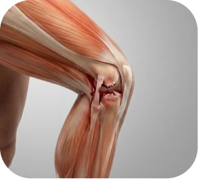
The posterior cruciate ligament (PCL) â the back-of-knee stabilizer â prevents the tibia (shin bone) from sliding backward on the femur (thigh bone). PCL tears occur through mechanisms that are distinctly different from ACL tears: a direct blow to the front of the tibia while the knee is bent, a car accident dashboard impact, or a hyperextension event.

The PCL is the strongest ligament in the knee â approximately twice the cross-sectional area of the ACL â and it provides the primary restraint against posterior tibial translation: the tibia sliding backward relative to the femur. When the PCL tears, this posterior restraint is lost. MRI characterizes the tear grade, the anatomy of what else was injured, and the concurrent findings that determine whether surgery is appropriate and what procedure is required.
Â
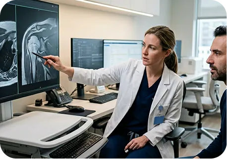
PCL tear MRI is interpreted across five key dimensions:
The PCL is graded using a three-tier system based on the degree of posterior tibial translation on clinical examination and stress imaging, combined with MRI findings.

A. Grade 1 â Mild Sprain (Almost Always Conservative)


The posterolateral corner (PLC) of the knee â comprising the lateral collateral ligament (LCL/FCL), popliteus tendon, popliteofibular ligament (PFL), and posterolateral capsule â provides the primary restraint against varus stress and external rotation. PLC injuries frequently accompany PCL tears, particularly in high-energy trauma and hyperextension mechanisms.
Â
Why PLC injury is the most important concurrent finding in PCL surgery:
Â
PLC components that require identification and surgical address:
Â


Chronic PCL deficiency â a PCL tear that has been present for months or years â produces characteristic secondary changes on MRI. The posterior tibial sag associated with PCL insufficiency overloads the medial compartment â particularly the medial femoral condyle and posterior horn of the medial meniscus. In chronic PCL deficiency, MRI frequently shows medial compartment cartilage thinning, medial compartment chondromalacia, and posterior medial meniscal tearing from the altered load distribution.
Â
When these secondary changes are identified on MRI in a chronic PCL-deficient knee, they provide additional urgency for surgical reconstruction â preventing further medial compartment deterioration that could eventually require knee replacement.


*Same-day consultations may be available

Unlike ACL tears â where surgery is generally recommended for active adults â the PCL surgery vs. conservative management decision is genuinely more nuanced. The PCL literature contains credible evidence for both surgical and non-operative management for selected Grade 3 tears. Understanding the factors that drive this decision is essential for any patient researching PCL treatment options.

The key distinction that separates surgical from non-surgical PCL management:


The published literature on conservative management of Grade 3 isolated PCL tears shows that while some patients achieve functional stability, a meaningful proportion develop progressive posterior instability, secondary medial compartment damage, and eventual reconstruction requirements anyway â just later and with more secondary damage accumulated â which is why at The Joint Preservation Center we present this evidence honestly and make the surgery vs. conservative decision at consultation with the full imaging and clinical picture, neither recommending surgery for patients who are appropriate candidates for conservative management nor withholding reconstruction from those who need it but remain uncertain.
For patients who have undergone appropriate conservative management â quadriceps rehabilitation, functional bracing, activity modification â and continue to experience posterior knee instability, the signals that reconstruction is appropriate are specific and recognizable.
Â
PCL treatment â from conservative rehabilitation for Grade 1-2 isolated tears to complex multiligament reconstruction for PCL + PLC + ACL injuries â spans a wider range than any other condition in this series. The appropriate procedure depends on tear grade, PLC involvement, concurrent ligament injuries, chronicity, patient age, activity level, and functional demands.
Indicated for Grade 1, Grade 2 isolated, and selected Grade 3 isolated PCL tears in appropriate patients (see Section 6). Core rehabilitation: aggressive quadriceps strengthening (open-chain leg extension), hamstring flexibility (hamstring pull produces posterior tibial translation in PCL-deficient knees), functional bracing, and progressive return to activity.
Â
Key principle of PCL conservative rehabilitation: the quadriceps is the primary dynamic substitute for the PCL â it pulls the tibia forward, countering the posterior sag. Quadriceps strengthening is the foundation of all PCL conservative and post-operative rehabilitation.

PCL reconstruction replaces the torn posterior cruciate ligament with a graft, placed through bone tunnels in the femur and tibia, that replicates the anatomic course and function of the native PCL. Unlike ACL reconstruction â where the transtibial technique is nearly universal â PCL reconstruction has two distinct tunneling approaches with different biomechanical implications.
In transtibial PCL reconstruction â the most commonly performed technique â the tibial tunnel is drilled from the front of the tibia upward and backward to the PCL’s posterior tibial attachment, creating a graft course from femur through the intercondylar notch to the posterior tibia; the key limitation of this approach is the “killer turn,” a sharp angular bend the graft must make at the posterior tibial cortex as it exits the tunnel, which creates stress concentration at the posterior tibial aperture and has historically contributed to higher graft failure rates in PCL reconstruction compared to ACL â a concern that modern improvements in graft positioning, fixation technique, and graft material selection have mitigated but not eliminated.
The tibial inlay technique addresses the killer turn problem by approaching the posterior tibia directly â through a posterior (back-of-knee) approach â and inlaying the graft directly into the posterior tibial attachment site. The tibial inlay technique eliminates the sharp angular bend at the posterior tibial cortex entirely.
Â
Advantages of tibial inlay: Eliminates graft stress concentration at the posterior tibial aperture. More anatomic graft course from femur to tibia. Preferred by many surgeons for isolated PCL reconstruction and for PCL combined with PLC where the posterior approach is already being used for PLC repair.
Â
The choice between transtibial and tibial inlay approaches is surgeon-dependent and based on the specific anatomy, concurrent injury pattern, and technical experience. Both produce reliable posterior stability when properly performed.

The PCL has two functional bundles â the dominant anterolateral (AL) and the posteromedial (PM) â that work together across the full range of knee flexion; single bundle reconstruction replaces the AL bundle and produces reliable functional stability for most patients, while double bundle reconstruction replaces both, providing more complete restoration of posterior tibial translation across the full range of motion at the cost of greater surgical complexity, longer operative time, and potential for over-constraint â making double bundle increasingly favored for high-demand athletes and cases where maximum posterior restraint restoration is the priority.

Unlike ACL reconstruction â where autograft is strongly preferred for younger athletes â PCL reconstruction more commonly uses allograft tissue, particularly for primary PCL reconstruction. The PCL is a large ligament requiring a large-diameter graft, and allograft avoids the additional morbidity of harvesting a large autograft.

Achilles tendon allograft: The most commonly used graft for PCL reconstruction. The Achilles allograft provides a large cross-sectional area appropriate for the PCL, with a bone plug that can be fixed in the femoral tunnel for secure bone-to-bone healing. For tibial inlay reconstruction, the bone block is inlaid directly into the posterior tibia.

Quadriceps tendon allograft or autograft: An alternative to Achilles allograft with favorable mechanical properties. Autograft quad tendon avoids donor tissue risk while providing a large graft appropriate for PCL reconstruction.

Hamstring tendon autograft (semitendinosus/gracilis): Used when autograft is preferred, particularly for younger patients where graft biology (ligamentization) may favor autograft over allograft. Multiple strands are folded to achieve adequate graft diameter.

Patellar tendon (BPTB): Occasionally used for PCL reconstruction â provides bone-to-bone fixation at both the femoral and tibial ends â but harvest site morbidity and graft size considerations make it less commonly chosen than Achilles allograft for primary PCL reconstruction.
When the posterolateral corner is injured alongside the PCL â the most surgically important concurrent finding â PLC reconstruction or repair is performed at the same surgical procedure. Failing to address the PLC when it is injured is the most common cause of PCL reconstruction failure.
The appropriate PLC procedure depends on the specific anatomic injury pattern, the acuity of the injury, and the tissue quality available for repair.

Primary PLC repair (acute injuries): When the PLC injury is acute (within 2â3 weeks) and the tissue quality is adequate, primary repair of the LCL/FCL, popliteus tendon, and popliteofibular ligament may be performed â restoring the native anatomy without graft harvest. Primary PLC repair requires timely surgery â delay beyond 2â3 weeks substantially reduces the prospects for primary repair.

PLC reconstruction with graft (chronic or poor tissue quality): When primary PLC repair is not feasible â due to chronicity, poor tissue quality, or the specific injury pattern â graft reconstruction of the key PLC structures (LCL, popliteus tendon, PFL) is performed using local tendon autograft or allograft. Multiple reconstruction techniques are available; the choice depends on which specific structures are injured.
When both the PCL and ACL are torn, both require surgical reconstruction. The complexity of bicruciate surgery typically necessitates staging â addressing one cruciate first and the second in a delayed procedure â or combined single-session surgery in selected cases. Bicruciate reconstruction is among the most technically demanding knee procedures and requires specialized multiligament expertise.

PCL reconstruction is more technically variable than ACL reconstruction â multiple approaches, graft options, and bundle configurations exist, each with specific advantages and indications. Understanding these options helps patients engage productively at consultation.
Transtibial reconstruction: The femoral and tibial tunnels are both drilled from the front â standard arthroscopic approach. The graft passes through both tunnels. The limitation: the graft must make a sharp turn at the posterior tibial cortex as it exits the tibial tunnel â the ‘killer turn’ â which creates graft stress concentration. Modern graft positioning and fixation improvements have reduced but not eliminated this concern.

Tibial inlay reconstruction: The tibial attachment is approached directly from the back of the knee through a posterior incision. The graft bone block is inlaid directly into the posterior tibial PCL footprint â creating a straight-line graft course without the killer turn bend. Most surgeons performing tibial inlay use it in combination with the posterior approach that is also used for concurrent PLC repair.
Transtibial reconstruction: The femoral and tibial tunnels are both drilled from the front â standard arthroscopic approach. The graft passes through both tunnels. The limitation: the graft must make a sharp turn at the posterior tibial cortex as it exits the tibial tunnel â the ‘killer turn’ â which creates graft stress concentration. Modern graft positioning and fixation improvements have reduced but not eliminated this concern.

Single bundle reconstruction: Reconstructs the dominant anterolateral (AL) bundle of the PCL â the primary restraint against posterior tibial translation at functional knee angles. Single bundle produces reliable posterior stability for most patients and is the most commonly performed PCL reconstruction technique.

Double bundle reconstruction: Adds reconstruction of the posteromedial (PM) bundle alongside the AL bundle, providing more complete posterior restraint through the full range of knee motion. More surgically complex, longer operative time, but increasingly favored for high-demand athletes and cases where maximum stability restoration is the priority.
High-energy PCL injuries â from football tackles, car accidents, wrestling, and direct blows â frequently involve concurrent damage to the posterolateral corner, ACL, MCL, menisci, and in severe cases, neurovascular structures. The PCL is rarely the only structure injured when the knee sustains the high energy required to tear the strongest ligament in the joint.
The PCL + PLC combined injury is the most surgically important pattern in posterior knee instability surgery. The combined posterior tibial translation (PCL) and external rotation/varus instability (PLC) creates a complex rotational-posterior instability that is functionally disabling and that cannot be managed conservatively in active patients.
Â
The PCL + PLC combination is diagnosed by:
Â
Surgical planning for PCL + PLC:

Both cruciate ligaments torn. Both require reconstruction. Staging vs. simultaneous reconstruction depends on the swelling and stiffness present, the concurrent injury pattern, and the surgeon’s assessment. Bicruciate injuries from high-energy trauma or knee dislocation equivalents require comprehensive multiligament planning.

A true knee dislocation â in which the tibia and femur completely separate â involves PCL tears in virtually 100% of cases alongside ACL, collateral ligament, and often PLC injuries. Knee dislocation is a vascular emergency before it is a surgical planning problem: popliteal artery injury occurs in 7â40% of knee dislocations and must be assessed with CT angiography or vascular Doppler before any ligament reconstruction proceeds. Peroneal nerve injury is also common.
Â
Multiligament reconstruction after knee dislocation is staged â acute neurovascular stabilization first, ligament reconstruction at an appropriate interval depending on swelling, neurovascular status, and the specific injury pattern. This is among the most complex surgery in knee reconstruction and requires specialized multiligament expertise.

Meniscal tears and articular cartilage damage are identified on MRI and addressed concurrently with PCL reconstruction when repair or treatment is indicated. As noted in Section 5, chronic PCL deficiency produces characteristic medial compartment overload â making the identification and treatment of medial compartment pathology an important component of PCL reconstruction planning in chronic cases.

Many PCL tears are initially managed conservatively â and some patients do well for years. But for a meaningful subset of PCL-deficient patients, the passage of time does not bring stability. Instead, the knee becomes increasingly unreliable, medial compartment pain develops, and activities that were manageable one year become limiting the next.
Â
Chronic PCL deficiency is not just the persistence of an old injury â it is an active process of progressive joint damage:
Â
PCL reconstruction in chronic PCL deficiency arrests the secondary damage cascade. Published series show that reconstruction in chronic PCL-deficient knees reduces medial compartment contact stress, addresses posterior tibial translation, and allows most patients to return to meaningful sport and physical activity â even when performed years after the original injury.
Â
The limitation in chronic cases: reconstruction does not reverse established articular cartilage damage or fully restore medial compartment health that has already deteriorated. Earlier reconstruction â while some cartilage health remains â produces better long-term outcomes than reconstruction performed after significant medial compartment arthritis has developed.
Â
For patients who have been managing a chronically unstable PCL for years, a consultation at The Joint Preservation Center will assess the current degree of posterior laxity, evaluate the status of the medial compartment and posterior meniscus, and determine whether reconstruction can still arrest the secondary damage cascade before it becomes irreversible.
PCL reconstruction failure â persistent posterior instability, graft elongation, or graft rupture â is more common than ACL reconstruction failure in the published literature, partly because of the killer turn graft angulation in transtibial techniques and partly because concurrent PLC injuries are sometimes missed at primary surgery.
The single most important reason for PCL reconstruction failure is unrecognized and unaddressed concurrent posterolateral corner injury at the time of primary PCL surgery. When the PLC is left unrepaired alongside a PCL reconstruction, the persistent rotational-varus instability from the PLC places the PCL graft under pathological loading during every weight-bearing activity. The graft stretches out (graft elongation) or ruptures.
Â
Before any revision PCL surgery, a meticulous assessment of PLC integrity is mandatory â clinical dial test examination and MRI characterization of the PLC components. Revision PCL reconstruction without concurrent PLC reconstruction (when indicated) is likely to fail again for the same reason.

Â
Second opinion before revision PCL surgery is strongly recommended. Understanding precisely why the primary reconstruction failed â which requires review of the original operative report, current MRI with dedicated PLC sequences, clinical dial test examination, and stress X-rays â is essential before committing to a revision approach.
PCL reconstruction recovery is generally comparable in duration to ACL reconstruction recovery â typically 9â12 months for full return to contact sport â but with a distinct rehabilitation emphasis. The quadriceps is the primary dynamic substitute for the PCL, and quadriceps strengthening is the foundation of all PCL rehabilitation. Crucially, hamstring exercises that pull the tibia backward (posterior tibial translation) must be carefully controlled in early recovery â the same direction as PCL-deficient knee instability â to protect the healing graft.
Phase 1: Immediate Post-Op (Weeks 0â4)
Extended knee brace locked in extension. The PCL heals with the knee in extension â the position that relaxes the PCL and allows optimal graft healing without posterior tibial sag. Early quad activation is critical. Crutches for protected weight bearing. Ice and elevation for swelling control.
Â
Phase 2: Progressive Motion (Weeks 4â10)
Gradual flexion restoration â progressive range of motion in a controlled hinged brace. Hamstring exercises are initially restricted. Open-chain quad exercises begin. Full weight bearing progresses.
Â
Phase 3: Strengthening (Weeks 10â20)
Progressive quadriceps strengthening â the foundation of PCL rehabilitation. Hamstring strengthening introduced carefully with attention to avoiding excessive posterior tibial force. Functional movement training begins.
Â
Phase 4: Sport-Specific Training (Months 5â9)
Sport-specific movement patterns. Running program progresses. Cutting, pivoting, and deceleration activities introduced progressively.
Â
Phase 5: Return to Sport (Months 9â12)
Return to full sport cleared based on objective criteria: quad and hamstring strength symmetry, hop test performance, and posterior stability assessment. Contact sport return at 9â12 months.

Three principles distinguish PCL rehabilitation from ACL rehabilitation:
Â
Drive?
Right leg surgery: typically 6â8 weeks. Left leg: often within 2â4 weeks.
Â
Return to desk work?
Often within 2â3 weeks for non-physical jobs.
Â
Return to physical work?
Light duty at 3â4 months. Full physical duty at 5â6 months.
Â
Return to running?
Straight-line running typically at 4â5 months.
Â
Return to non-contact sport?
Cutting and pivoting training at 5â6 months.
Â
Return to contact sport (football, wrestling, rugby)?
Typically 9â12 months.
When PLC reconstruction or repair is performed concurrently with PCL reconstruction, recovery is typically extended by 4â6 weeks compared to isolated PCL reconstruction. The PLC healing â particularly for primary PLC repair â requires a longer period of protected range of motion before progressive loading. Full contact sport return after combined PCL + PLC reconstruction is typically 12 months.
Weeks 1â2: Brace locked in extension. Crutches. Ice and elevation. Quad activation exercises immediately.
Â
Weeks 2â6: Brace unlocked for progressive flexion. Weight bearing progresses. Range of motion carefully advanced.
Â
Weeks 6â10: Full weight bearing. Progressive flexion continuing. Open-chain quad strengthening advancing.
Â
Months 3â4: Bike, pool running, progressive strength training. Daily activities fully managed.
Â
Months 4â6: Running program begins. Sport-specific strength and movement training.
Â
Months 6â9: Cutting and pivoting training. Non-contact sport participation begins.
Â
Months 9â12: Return to contact sport and full competition when objective milestones are met.

Modern PCL treatment is associated with:
Restored posterior knee stability
Significant improvements in pain and function
Improved functional outcome scores
High patient satisfaction
1
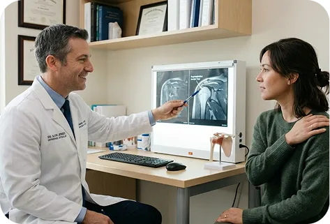
We evaluate your PCL tear grade, PLC status (clinical dial test + MRI), concurrent ligament injuries, chronicity, and functional instability. For acute injuries we provide same-day or same-week evaluation â particularly important for PCL + PLC injuries where the window for primary PLC repair closes at 2â3 weeks. For chronic patients we assess current posterior laxity, medial compartment status, and the secondary damage that conservative management has permitted to accumulate.
2
We present the evidence for conservative management vs. PCL reconstruction for your specific grade and injury pattern. For patients with PCL + PLC, combined PCL + ACL, or knee dislocation â the surgical recommendation is clear. For isolated Grade 3 PCL tears in selected patients, we present both options honestly. We recommend the most appropriate reconstruction technique â transtibial vs. tibial inlay, single vs. double bundle, graft selection â based on your injury pattern and functional demands.
3
If surgery is recommended, our fellowship-trained shoulder surgeons perform the appropriate procedure â PCL reconstruction (transtibial or tibial inlay), single or double bundle reconstruction, PLC repair or reconstruction when indicated, and concurrent ACL/MCL/meniscal procedures as needed â in a single surgical setting when feasible.
4
Our surgeons work closely with physical therapists to implement the PCL-specific rehabilitation protocol â with its emphasis on quadriceps strengthening, hamstring caution, and extended brace protocol â throughout the recovery phases.
5
Return to sport clearance is based on objective posterior stability assessment, strength symmetry testing, and functional testing â not calendar dates alone. The goal is a knee with restored posterior stability that can be trusted for sport, work, and daily function without bracing.
We track outcomes for five years after PCL reconstruction â monitoring posterior laxity, return-to-sport rates, medial compartment progression, PLC repair durability, and graft elongation rates by technique â so that our surgeons can continually refine technique selection, graft choice, PLC assessment protocols, and rehabilitation programs to achieve the most durable posterior stability outcomes over time.

1
Elite surgeons with decades of experience, incentivized to do the right thing
2
Prevent future surgeries
3
Heal with advanced, minimally invasive techniques
4
Preserve your natural joints, whenever possible
5
Seamless coordination from injury to recovery
6
Premium personalized care, made accessible
7
All patient outcomes tracked for 5 years
The posterior cruciate ligament (PCL) is the strongest ligament in the knee â it prevents the tibia (shin bone) from sliding backward relative to the femur (thigh bone). It runs from the back of the tibia to the inner (medial) surface of the lateral femoral condyle through the center of the knee. The ACL prevents the tibia from sliding forward; the PCL prevents it from sliding backward.
Â
PCL tears occur through different mechanisms than ACL tears: direct blow to the front of the tibia with the knee bent (dashboard injury, football tackle, wrestling), hyperextension events, and falls directly onto the knee. Unlike ACL tears â which almost always occur without direct contact â PCL tears most commonly require a direct force.
No â and this is one of the most important distinctions between PCL and ACL management. Grade 1 and Grade 2 isolated PCL tears are almost always managed conservatively with rehabilitation, and most do well. Even some Grade 3 isolated PCL tears in lower-demand patients can be managed conservatively with appropriate quadriceps rehabilitation.
Â
Surgery is strongly indicated when: the PCL is combined with a posterolateral corner (PLC) injury (almost always surgical), for Grade 3 PCL tears in competitive athletes and high-demand individuals, when conservative management fails, and when secondary medial compartment damage is developing. See Section 6 for the complete decision framework.
The posterolateral corner (PLC) is a complex of ligaments and tendons on the outer-back aspect of the knee â including the lateral collateral ligament (LCL/FCL), popliteus tendon, and popliteofibular ligament â that provide resistance to varus stress and external rotation. When the PLC is injured alongside the PCL, the combined posterior-rotational instability cannot be managed conservatively and almost always requires surgical repair.
Â
More importantly: a missed PLC injury is the most common cause of PCL reconstruction failure. When the PCL is reconstructed without addressing the concurrent PLC injury, the PCL graft fails under the ongoing rotational instability. Before any PCL reconstruction, PLC integrity must be carefully assessed clinically (dial test) and on MRI.
Both are PCL reconstruction techniques â the difference is how the tibial side of the graft is attached. Transtibial drilling creates a tibial tunnel from the front of the tibia, and the graft must make a sharp angular bend (‘killer turn’) at the posterior tibial cortex. Tibial inlay approaches the posterior tibial attachment directly from the back of the knee, placing the graft bone block directly into the tibial PCL footprint â eliminating the killer turn bend entirely.
Â
Both techniques produce reliable posterior stability in experienced hands. The tibial inlay is particularly appropriate when a posterior surgical approach is already being used for concurrent PLC repair. See Section 9 for the full technique comparison.
The PCL has two functional bundles â anterolateral (larger, dominant) and posteromedial. Single bundle reconstruction replaces the dominant anterolateral bundle â producing reliable functional stability for most patients. Double bundle reconstruction replaces both bundles â more anatomic and produces more complete restraint across the full range of motion, but is more technically complex. Double bundle is increasingly preferred for high-demand athletes.
A true knee dislocation occurs when the tibia and femur completely separate â tearing the PCL, ACL, and often multiple other ligaments simultaneously. Knee dislocation is a potential vascular emergency: the popliteal artery runs directly behind the knee and is injured in 7â40% of knee dislocations. If you have been told you dislocated your knee, or if your knee was significantly displaced during an injury, vascular assessment (CT angiography or Doppler imaging) is mandatory before any ligament reconstruction planning. Peroneal nerve injury is also common. Contact a specialist immediately.
Yes â and for PCL surgery in particular, given the nuanced surgery vs. conservative management decision for isolated Grade 3 tears, a second opinion from a fellowship-trained multiligament knee specialist is worthwhile. We offer second opinion consultations, including virtual consultations.
The Joint Preservation Center accepts most PPO insurance plans that have out-of-network benefits:
Â
If you have a PPO insurance plan with out-of-network benefits:
Â
Â
Note: If you have Medicare, Medicaid, TRICARE, or VA programs, or if your PPO does not have out-of-network benefits, you can still see our specialists and the surgery center will still be in-network. In this case, our specialists charge $250 for the initial office visit (all follow ups are included). Surgery is typically in the range of $6K â $8K depending on what you need done.
Â
If you are unsure whether your plan is accepted, our team can verify your coverage before your appointment.
*Same-day consultations may be available
*Online visits available