
*Online Consultations Available

*Online Consultations Available
1,000+ Shoulder Surgeries performed
Rehab-IntegratedâĻ
Care
Outcomes Tracked for 5 Years

The long head of the biceps tendon (LHB) attaches inside the shoulder joint and runs through the bicipital groove before connecting to the superior labrum, making it vulnerable to inflammation, degeneration, and mechanical instability. MRI and ultrasound describe biceps tendon pathology across five key dimensions, and understanding what your imaging shows helps explain why conservative care may not be resolving your pain and what a surgical approach can address.Â
The distinction between acute tendonitis and chronic tendinosis matters clinically â and surgically.


Medial subluxation of the long head of the biceps tendon out of the bicipital groove is a structural condition that is almost universally managed surgically. Unlike tendonitis â which may respond to conservative care in early stages â biceps subluxation reflects mechanical instability that does not self-correct.


The long head of the biceps tendon can sustain partial tears, either from chronic mechanical wear within the groove or from acute loading events.
Â

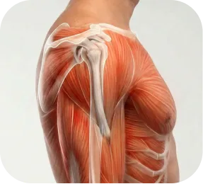
The bicipital groove is a narrow channel on the front of the humerus through which the LHB tendon travels. Its anatomy â width, depth, medial wall angle, and the presence of bone spurs â significantly affects whether the tendon can glide freely or becomes mechanically compressed.
Â

The LHB tendon does not exist in isolation. Biceps tendonitis and subluxation frequently occur alongside other shoulder conditions that MRI may identify simultaneously. The presence of concurrent pathology influences surgical planning â often allowing multiple structures to be addressed in a single arthroscopic procedure.


*Same-day consultations may be available


MRI and ultrasound provide critical structural information but do not establish a complete diagnosis on their own. Biceps tendon pathology must be interpreted alongside symptom pattern, clinical examination findings, occupational and athletic demands, and prior treatment history. A shoulder specialist integrates all of these factors before determining whether biceps tenodesis, tenotomy, or groove release is the most appropriate surgical approach for your situation.Â

Early-stage acute tendonitis may quiet down with rest, anti-inflammatory medication, and activity modification, but once the tendon has progressed to chronic tendinosis or subluxation from the groove, neither condition resolves without intervention. Degenerative changes within a chronically inflamed tendon do not reverse with conservative care, and a subluxating tendon will not return to a stable groove position without surgical relocation. For patients who have already tried physical therapy, cortisone injections, PRP, and prolonged rest without sustained improvement, this reflects the biological limits of conservative care once tendinosis or subluxation is established.Â
Refractory biceps tendonitis varies in its progression. Some patients maintain a tolerable level of discomfort with periodic injections; others experience gradual worsening despite ongoing treatment. The primary risks of leaving structurally significant pathology unaddressed include:
Â

As biceps tendon pathology progresses without treatment:
Â
In daily activity:
Â
Â
Occupational impact:
Â
Â
Athletic and sport impact:
Â
Â
For active adults in their 30s, 40s, and 50s â the demographic most commonly affected by surgical biceps tendonitis â delay often means extended time away from the training and occupational activities that define quality of life. Early surgical evaluation does not mean immediate surgery; it means understanding your options before the condition becomes more complex.
Most patients who reach a surgical evaluation for biceps tendonitis have already tried months of physical therapy, multiple cortisone or PRP injections, and activity modification without achieving lasting relief. This reflects a fundamental biological reality: once the long head of the biceps tendon has progressed to chronic tendinosis or is mechanically subluxating out of the bicipital groove, rehabilitation and injections cannot reverse that structural change. Conservative care can reduce inflammation around a degenerate tendon but cannot restore its structure or return it to the groove.Â
Conservative care CAN:
Â
Â
Conservative care CANNOT:
Â

Appropriate treatment for long head biceps tendonitis depends on the structural nature of the pathology (tendonitis vs. tendinosis vs. subluxation), the degree to which conservative care has been exhausted, the condition of the surrounding shoulder structures, and the patient’s age, activity level, and occupational demands.
For patients in the early or moderate stages of LHB tendonitis â without confirmed structural degeneration, subluxation, or significant partial tearing â structured physical therapy combined with injection management is the appropriate starting point.


Physical therapy alone may be less appropriate when:
Â

Biceps tenodesis is the preferred surgical treatment for most active patients with refractory LHB tendonitis, tendinosis, subluxation, or high-grade partial tearing. The procedure detaches the damaged portion of the biceps tendon from its anchor inside the shoulder joint and secures it to a new position on the humerus â eliminating the intra-articular pain source while preserving biceps function, cosmesis, and strength.
Biceps tenodesis is typically recommended for:
Â
Biceps tenodesis can be performed using two primary anatomical approaches. The choice between them is based on the clinical scenario, patient anatomy, and the surgeon’s judgment.

Suprapectoral (Arthroscopic) Tenodesis
The tendon is secured to the proximal humerus within or just below the bicipital groove, typically using an interference screw or cortical button fixation. Performed entirely arthroscopically through small keyhole incisions â no additional open incision is required. Well-suited for patients undergoing concurrent shoulder procedures (rotator cuff repair, SLAP repair, subacromial decompression) in the same arthroscopic setting.

Subpectoral (Open or Mini-Open) Tenodesis
The tendon is secured at a more distal position on the humerus, below the pectoralis major tendon insertion. Requires a small additional incision in the axillary fold. The subpectoral position removes the tendon from the groove entirely, potentially providing more reliable pain relief for patients with significant groove pathology, groove stenosis, or groove bone spurs. The subpectoral approach is often preferred for patients with isolated biceps tendonitis whose primary pain generator is the groove itself.
Both approaches use secure fixation â typically an interference screw threaded into a bone socket, a cortical button, or suture anchor fixation â to anchor the tendon to the humerus. The choice of approach and fixation method is determined during consultation based on your imaging, anatomy, and whether concurrent procedures are being performed.
Biceps tenodesis is performed as an outpatient (same-day) surgery under regional anesthesia with sedation or general anesthesia.
During the procedure:
The procedure typically takes 30â60 minutes for isolated tenodesis, and somewhat longer when combined with rotator cuff repair or other concurrent procedures.
Sling
Typically 4â6 weeks. The sling protects the tenodesis fixation while the tendon heals to the bone. Elbow flexion against resistance â which loads the biceps â is restricted during this period.
Â
Return to Desk Work
Often within 1â2 weeks, working with the arm in the sling or with minimal loading.
Â
Return to Overhead Work (Construction, Trades, Painting)
Typically 4â5 months. Return to full duty overhead occupational work requires complete tendon healing and progressive strength restoration.
Â
Return to Weightlifting / Gym Training
Lower body and core work resume progressively within 6â8 weeks. Light upper body activity resumes at approximately 3â4 months. Bench press, overhead press, and heavy loaded movements typically resume at 4â6 months, guided by strength milestones.
Â
Return to Overhead Sport (Pitching, Swimming, Volleyball)
Interval throwing programs typically begin at 4â5 months. Full return to competitive throwing or overhead sport generally occurs at 6â9 months depending on the sport and level of competition.
Â
Return to CrossFit
Kipping pull-ups, muscle-ups, and loaded overhead movements typically resume at 5â6 months. Bodyweight and lower-body movements resume progressively from 6â8 weeks.

Biceps tenotomy is a simpler arthroscopic procedure in which the long head of the biceps tendon is released from its attachment at the superior labrum without reattaching it elsewhere. The tendon retracts and the muscle shortens slightly. Tenotomy reliably eliminates intra-articular LHB pain and can be performed entirely arthroscopically through the same small incisions used for standard shoulder arthroscopy.
Biceps tenotomy is most appropriate for:
Â
After tenotomy, the released biceps tendon retracts and creates a visible bulge in the lower upper arm known as a Popeye deformity, occurring in approximately 40 to 70% of patients. For many, this is a cosmetic change only with well-preserved arm function. However, for patients concerned about arm appearance, active weightlifters, or those who require full supination strength for their sport or occupation, the cosmetic and functional implications make biceps tenodesis the preferred approach. The tenodesis vs. tenotomy decision is made collaboratively with each patient based on activity level, aesthetic concerns, and occupational demands.Â

Tenotomy has a faster recovery than tenodesis because no tendon-to-bone healing is required.
Â
Sling
1â2 weeks for comfort only
Â
Return to desk work
1â2 weeks
Â
Return to overhead work
6â10 weeks for most occupational tasks
Â
Return to sport
2â4 months depending on activity demands
Â
Bench press and upper body loading
Typically resumes at 6â8 weeks

When bicipital groove stenosis or groove sheath thickening is the primary pain generator â without significant tendon degeneration or subluxation â arthroscopic groove sheath release can decompress the tendon within its sheath, eliminating the mechanical impingement without requiring tenotomy or tenodesis.
Â
Groove release is less commonly performed as an isolated procedure; it is more often combined with tenodesis in patients with both groove stenosis and tendinosis or subluxation.
The most common question patients with refractory biceps tendonitis ask before surgery is: should I get a tenodesis or a tenotomy? Both procedures eliminate the intra-articular pain source. The right choice depends on several patient-specific factors.
After tenotomy, the released tendon retracts and the biceps muscle belly shifts distally â creating a visible bulge in the lower arm sometimes called a Popeye deformity, after the cartoon character’s exaggerated forearm appearance.
Â
Clinically, the Popeye deformity is primarily cosmetic. Most patients retain very good overall arm function. However:
Â
Â
Biceps tenodesis prevents the Popeye deformity by anchoring the tendon at a new position before it can retract, preserving arm contour and supination strength.

For patients who have decided on tenodesis, the next question is often about the approach â subpectoral or suprapectoral.
Â
Both approaches have strong evidence supporting reliable pain relief and functional recovery. The primary differences:
Â
Â
The approach is determined by your imaging findings, anatomy, concurrent procedures, and surgical judgment at the time of your consultation. At The Joint Preservation Center, the recommendation is individualized â not a fixed protocol applied to every patient.
Long head biceps tendonitis rarely exists in complete isolation. MRI frequently reveals concurrent pathology â rotator cuff tearing, SLAP tears, subacromial impingement, or subscapularis tearing â that requires simultaneous surgical management. Addressing only the biceps tendon while leaving other pathology untreated may result in incomplete pain relief and persistent shoulder dysfunction.
Â
At The Joint Preservation Center, we evaluate the full spectrum of shoulder pathology and perform all indicated procedures arthroscopically in a single operation where possible.
Rotator cuff tears, particularly subscapularis and supraspinatus tears, frequently coexist with LHB tendinopathy and subluxation. Because the subscapularis is the primary stabilizer of the biceps tendon in the groove, a subscapularis tear almost invariably destabilizes the LHB and leads to secondary tendinopathy. When both conditions are present, biceps tenodesis and rotator cuff repair are performed in the same arthroscopic procedure, eliminating the need for a second operation and addressing all pain sources simultaneously. Recovery follows the rotator cuff repair timeline.Â

In younger overhead athletes, particularly throwing athletes, the biceps anchor is susceptible to SLAP tears under the repetitive traction forces of throwing. When a SLAP tear coexists with LHB tendinopathy, the surgical decision between SLAP repair and biceps tenodesis is nuanced. In younger athletes with good labral tissue quality, SLAP repair preserves the native biceps anchor and may allow full return to overhead throwing. In older athletes or those with concurrent LHB tendinopathy, biceps tenodesis addresses both the labral and tendon pathology simultaneously with generally faster recovery. This decision is individualized based on age, sport level, and the specific tear pattern.Â

Subacromial bursitis and impingement frequently coexist with LHB tendinopathy. When both conditions are present and the patient has failed conservative care for both, biceps tenodesis and subacromial decompression (acromioplasty + bursectomy) can be performed in the same arthroscopic procedure. This is one of the most common combined shoulder procedures.

Subscapularis tears and LHB subluxation are tightly linked â the subscapularis is the primary restraint keeping the biceps tendon in the groove. When a subscapularis tear is present alongside LHB subluxation, subscapularis repair and biceps tenodesis are performed together. Addressing the subscapularis tear without stabilizing the biceps tendon â or vice versa â risks incomplete resolution of the mechanical instability.

A biceps tenodesis that does not heal â due to fixation failure, tendon pullout, or biological non-healing â can leave patients with persistent pain, loss of arm contour, and residual front-of-shoulder symptoms. Revision tenodesis may be considered when imaging confirms fixation failure and clinical examination identifies the LHB as the ongoing pain source.
Â
At The Joint Preservation Center, we evaluate revision cases with advanced imaging and a thorough assessment of what contributed to the initial failure â including fixation method, tendon quality, and rehabilitation adherence.

Recovery timelines differ meaningfully between biceps tenodesis and tenotomy, because tenodesis requires tendon-to-bone healing while tenotomy does not. Your surgeon will provide a specific protocol based on the procedure performed and any concurrent shoulder procedures.
Phase 1: Protection (Weeks 0â4/6)
Sling immobilization protects the tenodesis fixation. The arm is kept in a sling, and elbow flexion against resistance is restricted to prevent tensile load on the healing tendon-bone interface. Gentle pendulum exercises and passive shoulder range of motion begin early. The priority is protecting fixation while preventing shoulder stiffness.
Â
Phase 2: Active Motion (Weeks 4/6â8/10)
Active shoulder and elbow range of motion resumes progressively as healing allows. The shoulder begins moving under its own power. Daily activity tolerance increases noticeably during this phase.
Â
Phase 3: Strengthening (After ~10â12 Weeks)
Controlled progressive strengthening of the biceps, rotator cuff, and shoulder girdle begins once the tenodesis has biologically integrated into the bone. Supination and elbow flexion strengthening progress carefully. Full return to loaded activity is guided by strength milestones, not calendar dates alone.

Recovery after tenotomy is faster because no tendon-to-bone healing is required. Sling use is limited to 1â2 weeks for comfort. Active range of motion begins earlier. Strengthening can typically begin at 6â8 weeks. Most patients return to occupational and recreational activities within 2â4 months.

Drive?
Usually when out of the sling and off narcotic pain medication â typically 2â3 weeks for tenodesis, 1â2 weeks for tenotomy.
Â
Return to desk work?
Often within 1â2 weeks for both procedures, working with the arm in a sling or minimally loaded.
Â
Return to manual labor / overhead trades?
Approximately 4â5 months after tenodesis; 2â3 months after tenotomy. Full overhead occupational demands require strength milestones to be reached.
Â
Return to weightlifting / bench press?
Lower body and core resumes within 6â8 weeks. Bench press and upper body pressing typically resumes at 4â6 months after tenodesis; 6â10 weeks after tenotomy.
Â
Return to overhead sport / pitching?
Interval throwing typically begins at 4â5 months after tenodesis. Full return to competition generally requires 6â9 months.
Â
Return to CrossFit?
Bodyweight and lower-body movements resume progressively from 6â8 weeks. Loaded overhead and kipping movements typically resume at 5â6 months after tenodesis.
One of the most common concerns for working adults is minimizing income disruption and planning around professional obligations.
Â
Desk work and computer-based roles: Most patients return within 1â2 weeks. Remote work may allow an even earlier transition.
Â
Supervisory or client-facing roles: Return within 1â2 weeks is typical when physical lifting is not required.
Â
Construction, painting, electrical, plumbing, and overhead trades: Return to light duty is possible within 4â6 weeks for tenotomy and 8â12 weeks for tenodesis. Return to full overhead duty typically requires 4â5 months for tenodesis and 2â3 months for tenotomy.
Â
Workers’ Compensation: For patients with occupational biceps tendonitis, we provide work restriction documentation and return-to-work clearance letters as needed for employers, occupational health departments, and workers’ compensation case managers.
Â
Patients who plan ahead consistently report a smoother, less stressful early recovery.
Week 1: Sling at all times. Pain management with medication, icing, rest. Pendulum exercises begin. Most acute post-operative discomfort resolves significantly by day 5â7.
Â
Weeks 2â3: First post-operative visit. Passive shoulder range-of-motion exercises begin. Desk work resumes if comfortable. Elbow flexion against resistance still restricted.
Â
Weeks 4â6: Progressive passive and assisted motion. Sling discontinued for tenodesis at approximately week 4â6 depending on fixation security. Active elbow and shoulder motion begins.
Â
Weeks 6â10: Active range of motion well established. Daily activities noticeably easier. Shoulder and elbow begin gentle strengthening.
Â
Months 3â4: Progressive strengthening. Light upper body activity. Most patients return to light overhead occupational tasks.
Â
Months 4â6: Return to sport-specific and job-specific training. Bench press, overhead press, and throwing progressively resume according to strength milestones.
Â
Months 6â9: Full strength recovery. Return to competitive overhead sport and full-duty overhead work. Strength continues improving for up to 9â12 months.

Modern arthroscopic repair techniques are associated with:
High rates of pain improvement
Significant gains in strength
Improved functional outcome scores
High patient satisfaction
1
We evaluate your symptoms, shoulder strength and movement, clinical examination findings (Speed test, Yergason test, O’Brien test), and review your X-ray and MRI. We discuss your occupational demands, training history, and prior treatment. If imaging has not been obtained, MRI or ultrasound can be ordered.
2
We explain whether continued conservative management or surgical intervention is appropriate â and if surgery is recommended, whether biceps tenodesis, tenotomy, or a combined procedure best serves your specific anatomy, activity level, and goals.
3
If surgery is recommended, our fellowship-trained shoulder surgeons perform biceps tenodesis or tenotomy â and any concurrent indicated procedures â through small keyhole incisions to eliminate the pain source and restore shoulder function.
4
Our surgeons work closely with physical therapists to guide a structured rehabilitation program that protects the repair during healing and progressively restores strength, endurance, and sport-specific or job-specific function.
5
Rehabilitation progresses through objective strength milestones so you can confidently return to the activities that matter most â your training, your sport, your trade, or the daily tasks that biceps tendonitis has been limiting.
This long-term follow-up helps us understand how shoulders recover beyond the early healing period â including strength, function, and return to activity. These insights allow our surgeons to continually refine surgical planning and rehabilitation strategies to support durable shoulderâĻperformance over time.

1
Elite surgeons with decades of experience, incentivized to do the right thing
2
Prevent future surgeries
3
Heal with advanced, minimally invasive techniques
4
Preserve your natural joints, whenever possible
5
Seamless coordination from injury to recovery
6
Premium personalized care, made accessible
7
All patient outcomes tracked for 5 years
Biceps tendonitis of the shoulder refers to inflammation, degeneration, or structural instability of the long head of the biceps tendon (LHB) â the portion of the biceps muscle that runs through the shoulder joint and attaches at the top of the shoulder socket. The LHB travels through a narrow channel called the bicipital groove on the front of the upper arm bone before entering the joint, making it vulnerable to both inflammatory and mechanical problems.
Â
Common forms of LHB pathology include:
Â
Common symptoms include:
Â
Â
For weightlifters, the symptom is often most pronounced during the descent of a bench press or with overhead pressing. For overhead athletes, pain during the acceleration and deceleration phases of throwing is characteristic.
Diagnosis is made by an orthopedic shoulder specialist through:
Â
Biceps tendonitis of the shoulder is best evaluated and treated by an orthopedic surgeon who specializes in shoulder and sports medicine conditions. For patients considering surgery, a fellowship-trained shoulder surgeon â who has completed an additional year of advanced training specifically focused on shoulder conditions â has the deepest expertise in biceps tenodesis, tenotomy, and combined shoulder procedures.
Not necessarily â but surgery becomes appropriate when conservative care has been exhausted without sustained relief. Most patients benefit from a trial of physical therapy, cortisone injections, and activity modification before surgical evaluation.
Â
Surgery is typically recommended when:
Â
Both procedures address the long head of the biceps tendon as the source of pain â but they do so differently:
Â
The Popeye deformity is the visible change in arm contour that can occur after biceps tenotomy â the released tendon retracts and the biceps muscle belly shifts distally, creating a bulge in the lower portion of the upper arm. The name refers to the exaggerated forearm appearance of the cartoon character Popeye.
Â
Clinically, the Popeye deformity is primarily cosmetic. Most patients retain excellent arm function, and elbow flexion strength is generally well-preserved. Supination strength (rotating the forearm palm-up) may be mildly reduced by 10â20%.
Â
For patients with cosmetic concerns, active weightlifters, bodybuilders, or competitive athletes who rely on maximal supination strength, biceps tenodesis â which prevents the deformity by anchoring the tendon before it can retract â is the preferred approach.
Biceps tenodesis is a reliable procedure for appropriately selected patients with refractory LHB tendonitis, tendinosis, or subluxation. Patient satisfaction rates are high in appropriately selected candidates, particularly when the biceps tendon is confirmed as the primary pain source through clinical examination and diagnostic injection.
Â
Outcomes depend on:
Â
Recovery after biceps tenodesis is longer than after tenotomy because the tendon must heal biologically to the bone:
Â
Â
Recovery after biceps tenotomy is faster â sling for 1â2 weeks, return to overhead work at 2â3 months, return to sport at 2â4 months.
Yes â lower body and core work can resume progressively within 6â8 weeks of either procedure. Upper body loading resumes on a progressive schedule guided by healing milestones. Your rehabilitation program is designed to get you back to full training as efficiently and safely as possible.
Yes, and we encourage it. If you have been told you need biceps tenodesis or tenotomy and want to confirm that recommendation â or if you want to understand the tenodesis vs. tenotomy decision in more depth before committing â we offer second opinion consultations, including virtual consultations for patients who cannot travel.
The Joint Preservation Center accepts most PPO insurance plans that have out-of-network benefits:
Â
If you have a PPO insurance plan with out-of-network benefits:
Â
Â
Note: If you have Medicare, Medicaid, TRICARE, or VA programs, or if your PPO does not have out-of-network benefits, you can still see our specialists and the surgery center will still be in-network. In this case, our specialists charge $250 for the initial office visit (all follow ups are included). Surgery is typically in the range of $6K â $8K depending on what you need done.
Â
If you are unsure whether your plan is accepted, our team can verify your coverage before your appointment.
*Same-day consultations may be available
*Online visits available