
*Online Consultations Available

*Online Consultations Available
1,000+ Shoulder Surgeries performed
Rehab-IntegratedâĻ
Care
Outcomes Tracked for 5 Years


A high-grade MCL tear means that the medial collateral ligament â the primary ligament on the inner side of the knee that resists valgus (inward buckling) forces â has partially or completely ruptured from its femoral or tibial attachment, leaving the medial knee structurally unstable under load.
Â
MRI reports can describe a high-grade MCL tear across five factors:
The medial knee is a layered structure, and MRI may show injury to one or more components.

A. Superficial MCL (sMCL) â Complete Rupture

The deep MCL (also called the medial capsular ligament) lies beneath the superficial MCL and attaches directly to the medial meniscus. Deep MCL tears accompany superficial ruptures in high-energy injuries and contribute to medial instability. Combined superficial and deep MCL involvement increases the risk of secondary meniscus damage.


This refers to the degree of ligament disruption and how much medial structural integrity remains.
A. Grade 3 â Complete Rupture
B. High-Grade 2 Progressing to Complete
Near-complete tears with significant functional instability â sometimes presenting identically to Grade 3 on clinical examination. Surgical consideration depends on the degree of instability, associated injuries, and the patient’s activity demands.
C. Combined Grade 3 with Associated Ligament Injury
Location along the MCL determines the repair approach and healing potential.
A. Femoral-Sided (Proximal) Tear

Mechanism and timeline of injury determine whether primary repair is biologically feasible and which surgical strategy is most appropriate.
A. Acute High-Energy Traumatic Tear
High-grade MCL tears rarely occur in isolation. MRI commonly reveals concurrent knee pathology that significantly affects surgical planning.

A. ACL Tear (MCL + ACL â Most Common Combined Pattern)


*Same-day consultations may be available

Even though MRI provides valuable structural information, it does not establish a complete diagnosis â a high-grade MCL tear must be interpreted alongside the injury mechanism, the degree of medial joint opening on valgus stress testing, associated ligament injuries, and the functional demands placed on the knee both now and over time, all integrated by a knee ligament specialist through comprehensive evaluation before defining the most appropriate path forward.

Natural healing of a complete MCL rupture is unreliable for active patients with functional instability â unlike lower-grade sprains, a Grade 3 tear disrupts the full structural continuity of the ligament, and without the appropriate biological and mechanical environment the torn ends may scar in a lengthened or malpositioned state, leaving the medial knee structurally lax rather than properly healed; MCL avulsion injuries carry even lower healing capacity, as the ligament-to-bone interface cannot re-establish without surgical reattachment, making comprehensive evaluation by a knee ligament specialist essential to determine whether your specific tear pattern has healing potential or requires surgical intervention.

High-grade MCL tears vary in clinical course. Some patients manage daily activities with compensatory muscle function for a period; others experience immediate and severe functional instability that prevents return to sport or work.
Â
Persistent medial instability accelerates secondary joint damage. The medial meniscus â which is directly attached to the deep MCL â is particularly vulnerable to progressive injury when the medial side of the knee lacks its primary ligamentous restraint. Progressive meniscal wear, medial compartment cartilage deterioration, and cumulative bone bruising are the structural consequences of unaddressed Grade 3 laxity.
When high-grade MCL instability is left untreated, the following structural and functional changes develop:
Â
Â
These changes translate into functional limitations:
Â
In daily life:
Â
Â
Sports & performance:
Â
Â
As secondary damage accumulates and the window for primary repair closes, the reconstruction required becomes more complex and the probability of full return to sport decreases.
Appropriate treatment for a high-grade MCL tear depends on multiple factors, including the grade and location of the tear, the presence of associated ligament injuries, the chronicity of the injury, tissue quality, and the patient’s activity demands and goals.
For carefully selected patients with isolated Grade 3 MCL tears â particularly those who are lower-activity, older, or whose instability is functional rather than structural â a supervised rehabilitation program may be appropriate as an initial management approach. It is important, however, to understand what therapy can and cannot accomplish.
A well-designed rehabilitation program can:
Â
Â
Some patients experience meaningful improvement within 6â12 weeks when therapy is structured and progressive.

Physical therapy alone may be less appropriate when:
For working adults and athletes, the decision to pursue surgical treatment is often practical: the knee is no longer meeting the demands of sport, training, or daily activity. If instability persists after structured rehabilitation, this reflects the structural limits of conservative care â and surgical stabilization may be required to restore reliable function.
When a high-grade MCL tear produces functional instability, involves bony avulsion, or has failed conservative management, surgical repair reestablishes the structural integrity of the medial side of the knee.
MCL repair directly reattaches the torn ligament to its original bony attachment â the medial femoral epicondyle or tibial insertion â restoring native anatomy; when the MCL has avulsed with a bone fragment, the fragment is reduced and fixed with screws or suture anchors, and when the tear is mid-substance or tissue quality warrants additional support, an internal brace or suture tape may be used to protect the repair during early healing; primary MCL repair is most appropriate in the acute setting, within approximately 2â3 weeks of injury, when tissue quality is sufficient to hold fixation.

When the MCL is anatomically restored to its native attachment:
Â
Â
For most patients, successful repair results in:
Â
Â
Outcomes depend on the type and location of MCL injury, the presence of associated ligament tears, timing of repair, surgical technique, and adherence to structured rehabilitation.

MCL repair is performed through a medial knee incision that allows direct visualization and repair of the torn ligament structures.
Â
The procedure is typically performed under regional anesthesia with sedation or general anesthesia.
Â
During surgery, the surgeon:
A. Evaluates the Tear
For avulsion injuries, the MCL is reattached to its native footprint using suture anchors placed into the prepared bone surface. The anchors hold strong sutures that are passed through the ligament tissue and tied under appropriate tension â restoring the ligament to its correct anatomic position. For bony avulsions with a bone fragment, the fragment is reduced and fixed with a cortical screw or suture anchor.
At The Joint Preservation Center, fixation configuration and augmentation decisions are individualized based on tear pattern, tissue quality, and the patient’s functional demands.
Valgus stress is applied to confirm medial stability and appropriate ligament tension before wound closure.
Â
The procedure typically takes one to one and a half hours, depending on complexity and concurrent pathology. Healing occurs gradually over several months as the ligament integrates with the bony attachment.
Pain and Early Recovery After Surgery
Â
Most patients experience moderate discomfort in the first several days, managed with a combination of regional anesthesia, oral medication, and icing.
Â
A hinged knee brace is worn to protect the repair, typically for 6 weeks depending on the extent of injury and the procedure performed. The brace allows controlled range of motion while restricting valgus stress on the healing ligament.
Â
Crutches are typically used for 2â4 weeks to limit weight-bearing on the operative leg during early healing.
Â
Pain typically improves steadily over the first 1â2 weeks. Sleeping with the leg elevated is most comfortable during the early phase.
MCL repair heals in phases. Although the ligament is secured during surgery, biological integration to the bony attachment takes time â and protection during early recovery is essential.
Â
Recovery progresses in structured stages:
Â
Phase 1: Protection and Passive Motion (Weeks 0â6)
Gentle guided range-of-motion exercises begin early to prevent stiffness while protecting the repair. Weight-bearing is limited. The repaired ligament is not yet structurally strong.
Â
Phase 2: Progressive Weight-Bearing and Active Motion (Weeks 4â10)
You begin transitioning to full weight-bearing as healing progresses. Active range-of-motion exercises and early strengthening are introduced.
Â
Phase 3: Strengthening (After approximately 10â12 weeks)
Controlled strengthening begins once the MCL has biologically started to integrate with the bone. Quadriceps, hamstring, and hip abductor strength are rebuilt progressively.
Successful recovery depends on:
Â
Evidence shows that untreated high-grade MCL instability can:
Â
Â
These structural changes may reduce the effectiveness of surgery and increase reconstruction complexity later.
Â
This does not mean every Grade 3 MCL tear requires immediate surgery. It does mean that appropriate timing matters â particularly for avulsion injuries and combined ligament tears â when the window for primary repair is limited.
One of the most common concerns for working adults considering MCL surgery is timing: how to schedule the procedure around professional obligations, minimize income disruption, and plan for the weeks when full function is not yet restored.
Â
Desk work and computer-based roles:
Most patients return to desk work within 1â2 weeks, working with the leg elevated and using crutches initially. Full seated desk work is generally comfortable within 2â3 weeks.
Â
Supervisory, managerial, or client-facing roles:
If your work does not require significant walking or standing, return within 1â2 weeks is typical. Driving is usually possible once the brace is appropriately weaned and you are off narcotic medication, generally within 4â6 weeks.
Â
Manual labor, construction, trades, or physical occupations:
Return to light duty may be possible within 2â3 months. Return to full loading duties typically requires 4â6 months. Some employers offer modified duty during this transition.
Â
Work restrictions documentation:
We provide work restriction letters and return-to-work clearance documentation as needed for your employer or occupational health department.
Return-to-sport timelines depend on the demands of your activity, the complexity of the MCL repair or reconstruction, and your progress through rehabilitation milestones.
These timelines are approximations. Your specific return-to-sport plan is developed collaboratively between your surgeon and your physical therapist based on objective strength and stability milestones.
Preparing your home and support system before surgery helps the early recovery period go more smoothly. Because you will be in a brace and using crutches for several weeks, some advance planning makes a meaningful difference:
Â
Â
Patients who plan ahead consistently report a smoother, less stressful early recovery.
Recovery after MCL repair can vary depending on the extent of ligament injury, the presence of concurrent procedures, and the specific fixation used.
Â
While general timelines can be helpful for orientation, your recovery plan will be personalized. Your surgeon will guide you through a protocol tailored to your knee, your goals, and your lifestyle.
Â
Week 1:
Hinged brace and crutches. Pain management with medication, icing, and elevation. Gentle ankle pumps and quad sets. Most discomfort resolves significantly by day 5â7.
Â
Weeks 2â3:
First postoperative visit. Passive range-of-motion exercises with a therapist begin. Pain continues to improve. Desk work may resume if comfortable.
Â
Weeks 4â6:
Progressive transition to partial then full weight-bearing. Brace continues. Active motion and early strengthening begin.
Â
Weeks 6â10:
Brace is typically weaned as healing progresses. Transition to full active motion. Strengthening continues with increasing resistance.
Â
Weeks 10â16:
Progressive strengthening â quadriceps, hamstrings, hip abductors. Sport-specific movement patterns introduced for eligible patients.
Â
Months 4â5:
Progressive sport-specific or job-specific rehabilitation. Non-contact sport return for eligible patients.
Â
Months 5â7:
Contact sport return criteria assessment â strength, stability, and functional testing. Full contact clearance for most athletes.
Â
Months 6â12:
Continued strength gains. Full confidence with high-demand activities. Strength and stability can continue improving for up to a year.
Not every high-grade MCL tear can be addressed with primary repair alone. Chronic instability requiring graft reconstruction, MCL avulsion with bony injury, combined multiligament tears, and failed prior MCL surgery each present distinct challenges that require advanced techniques and specialized experience.
Â
At The Joint Preservation Center, we evaluate the full spectrum of medial knee pathology and offer the appropriate procedure for your specific injury â rather than a one-size-fits-all approach.
When MCL instability is chronic â lasting months to years after the original injury â the residual ligament tissue has typically degraded beyond the point where primary repair can provide durable stability, making reconstruction with a tendon graft the appropriate solution; the graft replaces the deficient MCL with healthy tissue anchored to the anatomic femoral and tibial attachment sites, most commonly using the gracilis or semitendinosus tendons harvested from the patient’s own knee (autograft) â expendable inner hamstring tendons that can be taken without functional deficit â with allograft serving as an alternative in revision cases or when autograft harvest is not preferred.
Â
Who Is a Candidate for MCL Reconstruction?
Â
MCL reconstruction may be appropriate for patients with chronic MCL instability lasting more than 6â8 weeks where tissue quality precludes primary repair, a failed prior MCL repair, significant attenuation and laxity without a repairable ligament end, or a chronic combined injury pattern requiring multiligament reconstruction.
Â
MCL Reconstruction Recovery for Active Adults
Â
The hinged knee brace is worn for 6 weeks, with gradual range-of-motion and weight-bearing progression beginning in the early postoperative period. Active strengthening begins around weeks 10â12. Return to contact sport typically requires 6â9 months with objective strength milestone confirmation before clearance.
When the MCL tears away from its bony attachment â at the femoral origin or tibial insertion, with or without a bone fragment â avulsion fixation directly reattaches the ligament-bone unit to its native footprint: bony avulsions are reduced and held with a cortical screw or compression fixation while the soft tissue structures are repaired with suture anchors, pure soft-tissue avulsions are secured with suture anchors placed into the freshened bone surface, and when tissue quality is borderline an internal brace or suture tape is added to reinforce the fixation.
Â
Who Is a Candidate for Avulsion Fixation?
Â
Avulsion fixation is appropriate when MRI or X-ray confirms MCL detachment from its femoral or tibial attachment, the injury is in the acute or subacute phase (within approximately 4â6 weeks), and the avulsed tissue remains identifiable and suitable for reattachment. Stener-like lesions â where soft tissue is interposed and prevents spontaneous healing â are strong indications for surgical fixation regardless of timing.
Â
Recovery
Â
Recovery following MCL avulsion fixation follows a similar phased protocol to standard MCL repair, with the brace and weight-bearing timeline adjusted for bone healing at the fixation site. CT or X-ray confirmation of fragment consolidation guides progression to strengthening.
High-grade MCL tears frequently occur alongside other structural knee injuries. Addressing only the MCL when concurrent pathology is present may leave residual instability or accelerate secondary joint damage.
MCL Repair with ACL Reconstruction
MCL + ACL is the most common combined ligament injury in contact sports â typically from a valgus blow to a partially flexed, planted knee. When both ligaments are compromised, addressing the MCL alone leaves the ACL-deficient knee at risk for persistent anterior instability. We evaluate each case to determine the optimal staging (simultaneous vs. staged) and sequencing based on swelling, range of motion, and the specific injury pattern.
MCL Repair with Medial Meniscus Repair
The deep MCL attaches directly to the medial meniscus. When high-grade MCL tears include medial meniscal injury â peripheral detachment or posterior root involvement â concurrent meniscal repair at the time of MCL surgery preserves meniscal function and protects long-term joint health
MCL Repair with PCL or Posterolateral Corner Reconstruction
Combined medial and posterior or lateral knee instability â typically from very high-energy mechanisms including near-dislocations â requires multiligament reconstruction. These complex cases demand subspecialty experience in surgical sequencing, graft selection, and rehabilitation coordination.
Some patients present with MCL instability in both knees â particularly athletes with bilateral valgus stress exposure or patients with underlying ligamentous laxity. Bilateral involvement is managed with a staged approach: one knee is stabilized first, and the second is addressed after the first has recovered sufficiently to bear weight and manage daily activities.
Â
We develop a surgical and rehabilitation plan that sequences bilateral procedures to minimize cumulative downtime, which is especially important for working adults managing career obligations and caregiving responsibilities.
An MCL repair or reconstruction that does not hold â whether due to anchor pullout, graft elongation, technical failure, or a new traumatic event â leaves patients with persistent instability after already making the recovery investment. For working adults and athletes, a failed primary MCL procedure is especially disruptive.
Â
When Is Revision Surgery Appropriate?
Â
Revision MCL surgery is considered when imaging confirms structural failure, medial instability persists after adequate rehabilitation, and the remaining anatomy supports a revision procedure â assessed at The Joint Preservation Center with MRI and stress X-rays to determine current structural status and the cause of initial failure.
Â
Signs of Failure to Watch For
Â
Key warning signs include return of valgus giving-way after initial improvement, inability to progress through rehabilitation milestones, a re-injury event with immediate loss of medial stability, or recurrent instability in the months following primary surgery â any of which warrant prompt reevaluation.
Â
Revision Approaches
Â
Depending on the scenario, revision options include conversion to MCL reconstruction with a new graft (the most common pathway after failed primary repair), revision reconstruction with allograft when autograft is unavailable, or augmented fixation to address anchor pullout or bone loss at the attachment site â with the specific plan determined by what the anatomy and biology can support at reevaluation.
Chronic medial instability arthropathy is a distinct condition that can develop when longstanding, unaddressed MCL laxity leads to progressive medial compartment deterioration. Over time, the abnormal valgus load distribution â concentrated on the medial meniscus and articular cartilage â produces accelerated medial compartment wear, meniscal damage, and eventual joint-line narrowing. This is not simply an unstable knee â it represents a structural transformation of the medial compartment that requires a different management approach.
Â
When Joint Preservation Is the Right Option
Â
For patients with medial instability arthropathy, the priority is to address the underlying ligamentous cause â MCL reconstruction to eliminate valgus laxity â before medial compartment deterioration becomes irreversible, with concurrent meniscal pathology addressed at the same time to preserve native joint anatomy; for working-age patients and athletes, this decision carries particular weight, and recovery timelines, activity progression, and long-term durability are discussed in detail at consultation so you can make a fully informed decision about your knee’s trajectory.

Modern MCL repair and reconstruction techniques are associated with:
Restored medial knee stability
Significant gains in strength and range of motion
Improved functional outcome scores
High patient satisfaction
1
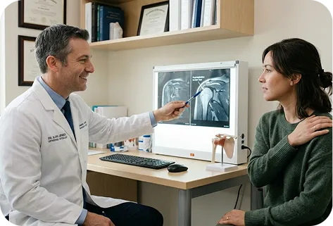
We evaluate your symptoms, medial knee stability, strength and range of motion, review your imaging â including MRI, stress X-rays, and CT where indicated â and consider your injury mechanism, functional demands, and goals. If additional imaging is needed, it can be ordered.
2
We explain whether rehabilitation or surgical stabilization is appropriate based on your MCL grade, tear location and pattern, tissue quality, the presence of associated injuries, and your activity goals.
3
If surgery is recommended, our fellowship-trained knee surgeons perform the appropriate procedure â primary MCL repair, avulsion fixation, MCL reconstruction with graft, combined ACL + MCL surgery, or revision stabilization â to restore medial knee stability and prevent further damage.
4
Our surgeons work closely with physical therapists to guide a structured rehabilitation program that restores motion and strength while protecting the repair or reconstruction.
5
Rehabilitation progresses through milestones so you can confidently return to the activities that matter most â work, exercise, and sport.
This long-term follow-up helps us understand how shoulders recover beyond the early healing period â including strength, function, return to sport, and return to throwing. These insights allow our surgeons to continually refine surgical planning and rehabilitation strategies to support durable shoulder performance over time.

1
Elite surgeons with decades of experience, incentivized to do the right thing
2
Prevent future surgeries
3
Heal with advanced, minimally invasive techniques
4
Preserve your natural joints, whenever possible
5
Seamless coordination from injury to recovery
6
Premium personalized care, made accessible
7
All patient outcomes tracked for 5 years
The medial collateral ligament (MCL) is the primary ligament on the inner side of the knee that resists valgus forces â the inward buckling of the knee under load.
Â
The MCL works to:
Â
A high-grade MCL tear â Grade 3 complete rupture or avulsion â means the ligament has lost its structural continuity, leaving the medial side of the knee without its primary stability restraint. The knee is structurally vulnerable to inward buckling under any valgus load.
High-grade MCL tears and medial knee instability can cause several symptoms depending on the severity and chronicity of the injury.
Â
Common symptoms include:
Â
Some patients experience immediate severe instability after a single traumatic event; others notice gradual progression of medial giving-way over time.
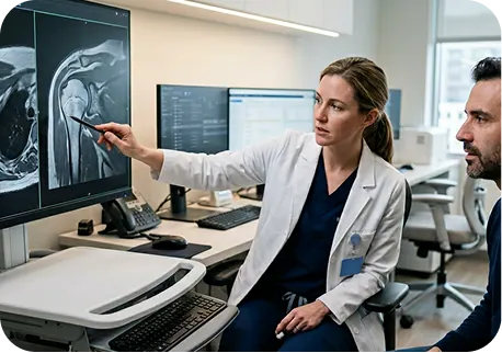
High-grade MCL tears are typically diagnosed by an orthopedic surgeon who specializes in knee ligament injuries.
Â
During evaluation, the surgeon reviews your injury mechanism and performs a physical examination that includes specific instability tests â the valgus stress test at 0° and 30° of flexion â to grade the degree of medial joint opening and confirm the structural loss.
Â
Imaging studies are then used to confirm the diagnosis and characterize the injury:
Â
Because imaging findings must be interpreted alongside clinical history and stress examination, the final diagnosis is based on a combination of all three.
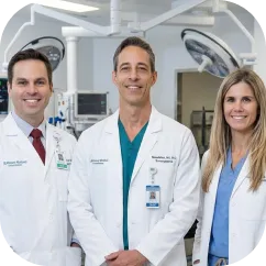
High-grade MCL tears are treated by orthopedic surgeons who specialize in knee ligament and sports medicine conditions.
Â
For the highest level of specialization, look for a fellowship-trained knee surgeon. Fellowship training is an additional year of advanced surgical training completed after orthopedic residency â focused specifically on complex knee ligament conditions and procedures.
Â
This training typically includes:
Â
Fellowship-trained knee surgeons often have deeper experience in the full spectrum of MCL procedures â from acute primary repair to complex chronic reconstruction â which matters most in cases involving avulsion, combined ligament tears, or prior failed surgery.
Patients do not feel pain during MCL surgery. TMCL surgery is performed under regional anesthesia with sedation or general anesthesia, so patients feel no pain during the procedure; afterward, some discomfort is expected during the first 48â72 hours and is managed with medication, ice, brace support, and elevation â often supplemented by a regional nerve block that keeps the knee numb for several hours post-operatively; pain then improves steadily as healing progresses, and for most patients the short-term recovery is worthwhile because successful stabilization eliminates giving-way episodes, restores knee confidence, and allows return to sport and work.
MCL repair and reconstruction are reliable procedures for appropriately selected patients, especially when the procedure matches the injury pattern and rehabilitation is followed closely.
Â
Outcomes vary with:
Â
In general, primary MCL repair for acute Grade 3 avulsion injuries achieves high rates of stability restoration. MCL reconstruction for chronic instability achieves reliable medial stability in the majority of patients, with return to sport rates comparable to ACL reconstruction outcomes in the published literature.
Â
At The Joint Preservation Center, our surgeons track outcomes for 5 years because durable medial knee stability â not just early pain relief â is the true measure of success.
Recovery after MCL surgery occurs in phases as the ligament heals to its bony attachment or the graft integrates biologically â most patients wear a hinged knee brace and use crutches for 4â6 weeks while guided motion is introduced to maintain mobility without stressing the repair, then progress to strengthening over the following weeks; meaningful day-to-day function typically returns within 3â4 months, while full confidence with contact sport, physical work, and high-demand activity requires 5â7 months for primary MCL repair and 6â9 months for reconstruction, with adherence to the brace protocol and a structured rehabilitation program playing a critical role in achieving the best possible outcome.
Unlike MCL repair and reconstruction â which restores native ligament anatomy and preserves the natural knee joint â knee replacement involves implanting metal and polyethylene components. Replacement is not a treatment for MCL instability or ligament tears.
Â
Â
Knee replacement is relevant only in very specific end-stage scenarios â such as medial compartment arthritis developing after decades of untreated MCL instability, which is precisely the outcome that early MCL stabilization aims to prevent.
Yes, many patients continue to use their knee with a known high-grade MCL tear, as the quadriceps and hamstrings can temporarily compensate for the structural deficiency â but this often comes with apprehension, avoidance of valgus-loading positions, and reduced confidence, and over time continued instability can lead to progressive medial meniscus injury, increasing cartilage wear, and eventual loss of functional capacity, which is why evaluation and treatment planning are important even when function feels manageable in the short term.
High-grade MCL tears most commonly occur from valgus force applied to the knee â the inner side of the knee being pushed inward while the outer side is blocked or planted.
Â
Contact sport mechanisms
are the most common cause â a tackle in football, a direct blow in soccer, a collision in rugby or hockey, a fall in wrestling or skiing with a valgus-forced knee position. The force drives the inner knee apart, rupturing the MCL fibers. A single high-energy impact can produce a complete Grade 3 rupture with avulsion.
Â
Non-contact valgus stress
from rapid deceleration, change-of-direction movements, or landing with the knee in valgus can also produce high-grade MCL injury â particularly in combination with ACL tears in cutting sports.
Â
Both patterns can result in progressive medial instability that limits sport, work, and daily activity â particularly when the initial injury is not definitively addressed.
Chronic MCL instability â persistent medial giving-way or valgus laxity despite conservative treatment â develops when a Grade 3 tear heals in a lengthened or attenuated state, or fails to heal adequately, and is an important clinical finding because it typically indicates degraded ligament tissue that precludes primary repair alongside progressive secondary damage to the medial meniscus and articular cartilage; graft reconstruction is the appropriate surgical solution in this setting, and evaluation by a knee ligament specialist experienced in chronic MCL reconstruction is recommended for any patient experiencing persistent medial instability despite conservative management.
Yes. It is common for MRI to reveal concurrent pathology alongside a high-grade MCL tear â including ACL tears requiring reconstruction, medial meniscus injury, PCL involvement, posterolateral corner instability, or bone bruising with chondral injury.
Â
We routinely address combined knee pathology during a single procedure or in a carefully planned staged approach. For example, MCL repair can be combined with ACL reconstruction, medial meniscus repair, or concurrent ligament reconstruction depending on what your imaging and clinical examination indicate. Addressing all contributing structures optimally avoids the need for additional surgery and typically produces better overall outcomes.
Bilateral MCL instability is managed with a staged approach â one knee is stabilized first, and the second is addressed after the first has recovered sufficiently to bear weight and manage daily activities comfortably. We develop a sequencing plan that minimizes total downtime, which is especially important for patients managing work obligations and family responsibilities.
Â
The Joint Preservation Center accepts most PPO insurance plans that have out-of-network benefits:
Â
If you have a PPO insurance plan with out-of-network benefits:
Â
Â
Note: If you have Medicare, Medicaid, TRICARE, or VA programs, or if your PPO does not have out-of-network benefits, you can still see our specialists and the surgery center will still be in-network. In this case, our specialists charge $250 for the initial office visit (all follow ups are included). Surgery is typically in the range of $6K â $8K depending on what you need done.
Â
If you are unsure whether your plan is accepted, our team can verify your coverage before your appointment.
*Same-day consultations may be available
*Online visits available