

*Online Consultations Available
*Online Consultations Available
1,000+ Knee Surgeries performed
Rehab-IntegratedâĻ
Care
Outcomes Tracked for 5 Years


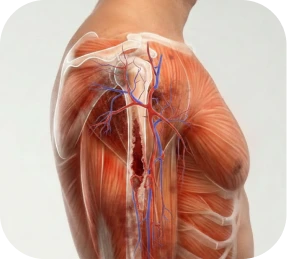
Shoulder fractures â broken bones in the proximal humerus (the ball of the shoulder), glenoid (the socket), or scapula (the shoulder blade) â are common injuries following high-energy trauma. Motor vehicle accidents, sports collisions, workplace accidents, and high-impact falls are the most frequent causes in working-age adults.
Â
The first 24â72 hours after a shoulder fracture are critical. Not every fracture requires surgery â but displaced fractures, multi-fragment fractures, fracture-dislocations, and fractures with nerve or vascular involvement require urgent surgical evaluation to determine whether early intervention produces the best outcome. Waiting weeks while swelling and the fracture pattern evolve can complicate surgical options.
A shoulder fracture â technically a fracture of the proximal humerus, glenoid, or scapula â must be thoroughly characterized by imaging before any surgical plan can be made. X-ray provides the initial diagnosis. CT scanning â particularly 3D reconstruction â is frequently essential for complex fractures, providing the three-dimensional detail needed to plan ORIF, arthroplasty, or glenoid/scapula reconstruction.
Â
Shoulder fracture imaging is evaluated across six key dimensions that directly determine whether surgery is indicated, which procedure is appropriate, and what approach and hardware will be used:
A. Proximal Humerus (Most Common)
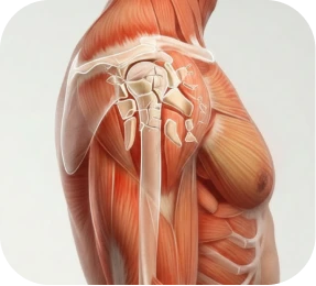
The ball of the shoulder joint. The proximal humerus is composed of four structural segments: the humeral head, the greater tuberosity, the lesser tuberosity, and the humeral shaft. The number of segments involved and their displacement defines the Neer classification â the most widely used system for surgical planning. Proximal humerus fractures account for the majority of shoulder fractures in working-age adults.

The bony prominence on the outer aspect of the proximal humerus where the supraspinatus and infraspinatus rotator cuff tendons attach. Displaced greater tuberosity fractures â particularly those displaced more than 5 mm â almost always require surgical fixation to restore rotator cuff function and prevent malunion with impingement.

The anterior prominence where the subscapularis attaches. Lesser tuberosity fractures are less common in isolation but clinically significant â a displaced fragment can destabilize the biceps tendon groove. Requires surgical reattachment when displaced.

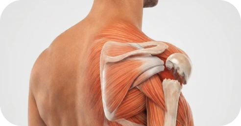
The socket side of the shoulder joint. Glenoid fractures can involve the rim (associated with dislocation â bony Bankart lesion), the articular fossa (intra-articular fractures requiring anatomic restoration), or the glenoid neck. Surgical fixation is indicated for displaced articular fractures and glenoid rim fractures causing instability.

The shoulder blade. Most scapular fractures heal non-operatively, but displaced fractures of the scapular body, neck, or glenoid face â and fractures combined with clavicle fractures (floating shoulder) â may require ORIF.
The Neer classification describes proximal humerus fractures by the number of displaced segments involved. A segment is defined as displaced if it has moved more than 1 cm or angulated more than 45 degrees from its normal position. This classification is the primary driver of the ORIF vs. arthroplasty decision.
A. One-Part (Non-Displaced)
All four segments remain in acceptable alignment regardless of fracture lines. Non-surgical management with a sling is typically appropriate. This category is NOT the target of this campaign â non-displaced fractures are conservative management.
One of the four segments is displaced. The surgical neck (shaft separating from the head), greater tuberosity, or lesser tuberosity may be the displaced component. Two-part surgical neck fractures with significant displacement in young active adults are typically treated with ORIF. Two-part greater tuberosity fractures displaced more than 5 mm require surgical fixation to restore rotator cuff function.
Two segments are displaced. The humeral head, one tuberosity, and the shaft are involved. Three-part fractures in active working-age adults with good bone quality are typically managed with ORIF using a locking plate or intramedullary nail. The goal is anatomic restoration of the joint to allow full recovery of strength and motion.
All four segments are displaced. The humeral head is completely detached from both tuberosities and the shaft. Four-part fractures carry the highest risk of avascular necrosis (AVN) of the humeral head â disruption of blood supply during the fracture may cause the bone to die despite successful surgical fixation. In younger, active patients with good bone quality, ORIF is attempted; in patients where the blood supply to the head is compromised or bone quality is poor, hemiarthroplasty or reverse shoulder arthroplasty for fracture may be recommended.
The shoulder blade. Most scapular fractures heal non-operatively, but displaced fractures of the scapular body, neck, or glenoid face â and fractures combined with clavicle fractures (floating shoulder) â may require ORIF.

Even within a given Neer classification, the degree of displacement and angulation affects surgical urgency and the complexity of fixation.
A. Minimally Displaced
Fragments are in near-anatomic position. Conservative management may be appropriate depending on the specific pattern and patient demands.
B. Significantly Displaced
Fragments have separated by more than 1 cm or angulated more than 45 degrees. Surgical fixation is generally indicated for displaced fragments in active working-age adults, because malunion or nonunion in the displaced position leads to predictable functional loss.
C. Severely Displaced / Unstable
Major displacement with no bony contact between fragments. Unstable patterns â where fragments continue to move with gravity â require early surgical stabilization. Secondary displacement during conservative management (the fracture shifts further out of position while in a sling) is a clear indication for surgical intervention.
Â
Comminuted fractures â in which the bone has fractured into multiple fragments rather than two clean pieces â are among the most challenging shoulder injuries to treat.
A. Simple (2 Fragments)
Clean fracture line between two major fragments. ORIF is typically straightforward with a locking plate or intramedullary nail.
Multiple bone fragments. Requires careful pre-operative planning, often with 3D CT reconstruction. Locking plate ORIF can accommodate comminuted patterns when sufficient bone quality allows secure fixation. When comminution is severe and the humeral head fragments cannot be reliably reconstructed, arthroplasty may be indicated.
The bone has fragmented into many pieces â sometimes described by patients as ‘my shoulder was completely shattered.’ Severely comminuted proximal humerus fractures in younger patients represent some of the most complex cases in shoulder surgery, requiring extensive pre-operative planning and sometimes intraoperative decision-making about whether ORIF or arthroplasty will produce the better long-term outcome.

Shoulder fractures â particularly high-energy fractures â frequently occur alongside other injuries that significantly affect surgical planning and urgency.

A. Fragments)Fracture-Dislocation
When a shoulder fracture occurs simultaneously with a shoulder dislocation, the combined injury is more complex and more urgent than either alone. Fracture-dislocations may involve the proximal humerus, the glenoid, or both â and the combined instability prevents simple sling management. Prompt reduction of the dislocation (repositioning the shoulder into the socket) is typically required, followed by surgical planning for fracture fixation. Locked fracture-dislocations â where the humeral head is locked in a dislocated position and cannot be closed-reduced â require urgent surgical intervention.

A posterior shoulder dislocation can impinge the soft anterior humeral head against the posterior glenoid, creating an impression fracture of the humeral head â the reverse Hill-Sachs lesion. When the impression fracture is large (involving more than 20â40% of the humeral head articular surface), it creates chronic instability if left untreated. Surgical options include the McLaughlin procedure (transferring the subscapularis into the defect), humeral head reconstruction, or replacement.
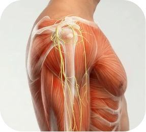
The axillary nerve â which powers the deltoid muscle and provides sensation to the outer arm â runs directly adjacent to the proximal humerus and is the most commonly injured nerve in proximal humerus fractures. Axillary nerve injury may cause arm weakness and deltoid paralysis. While many axillary nerve injuries recover spontaneously with time, severe injuries may require surgical exploration. The presence of axillary nerve symptoms warrants prompt neurological assessment.
High-energy shoulder fractures â particularly fracture-dislocations â can injure the axillary artery or its branches. Signs of vascular injury (cold, pale, or pulseless hand; rapidly expanding hematoma) constitute a surgical emergency requiring immediate vascular surgery consultation alongside orthopedic management.

Greater tuberosity fractures frequently involve concurrent rotator cuff tearing, as the fragment carries the supraspinatus and infraspinatus tendon attachments. When the greater tuberosity is repaired, concurrent cuff pathology is addressed at the same procedure.
Whether the fracture involves the articular cartilage surface of the humeral head or glenoid directly affects the surgical approach and long-term joint prognosis.

A. Extra-Articular
The fracture line does not cross the joint surface. Anatomic fixation can restore normal joint mechanics if achieved.

The fracture extends into the articular surface. Intra-articular fractures require anatomic restoration of the joint surface â any residual step-off of more than 2 mm in the glenoid or significant humeral head fragmentation is associated with post-traumatic arthritis. CT scanning is essential to characterize articular involvement and plan the surgical approach.
The humeral head itself is split into multiple fragments. This pattern â uncommon but highly complex â often requires humeral head replacement (hemiarthroplasty or RSA for fracture) rather than fixation, as the fragments cannot be reliably reassembled.

*Same-day consultations may be available


Plain X-ray confirms the fracture and provides initial classification. CT scanning â especially with 3D reconstruction â is the standard of care for complex proximal humerus fractures, glenoid fractures, and scapular fractures before surgical planning. CT allows precise characterization of fragment size, displacement, comminution, articular involvement, and bone quality that plain X-ray cannot provide.
Â
At The Joint Preservation Center, pre-operative CT is routinely used for all complex shoulder fractures to optimize surgical planning and implant selection before the patient enters the operating room.

Not every shoulder fracture heals as expected. Two distinct problems can develop weeks to months â or even years â after the initial injury:
A shoulder fracture nonunion occurs when the broken bone fails to heal after an appropriate period of time â typically defined as six months or longer without radiographic evidence of healing. The fracture site remains a mobile fibrous junction rather than solid bone, producing persistent pain, weakness, and functional limitation.
Nonunions can develop for several reasons:
Â
Â
Nonunions are classified by their radiographic appearance:
Â
Â
Surgical treatment for shoulder nonunion typically involves: revision fixation with a locking plate or intramedullary nail, bone grafting (autograft from the patient’s own bone or allograft) to restore the biological environment, and in cases where the fracture fragments are not salvageable, conversion to shoulder arthroplasty.

A shoulder malunion occurs when the fracture heals â but in a displaced or angulated position rather than the correct anatomic alignment. Malunions are common when displaced fractures are managed non-operatively, or when initial surgical fixation does not achieve or maintain anatomic reduction.
The consequences of shoulder malunion depend on the location and degree of malalignment:
Â
Â
Surgical treatment for shoulder malunion ranges from corrective osteotomy (surgically re-breaking and realigning the bone) for isolated tuberosity malunions to shoulder arthroplasty for complex malunions with post-traumatic arthritis or AVN.

Not all shoulder fractures require surgery. Non-displaced and minimally displaced fractures in appropriate patients can heal successfully with sling immobilization and supervised rehabilitation. When this approach is appropriate and followed correctly, it produces good outcomes.
Â
However, conservative management has specific limits â and recognizing those limits is critical to avoiding preventable complications: nonunion, malunion, post-traumatic arthritis, and persistent functional loss.
Â
For working-age adults whose livelihood depends on shoulder function â construction workers, tradespeople, athletes, first responders â the threshold for surgical consultation is lower. The functional and economic consequences of prolonged non-healing or malunion in this population make early specialist evaluation a practical necessity.

Shoulder fracture treatment is individualized based on fracture type, location, displacement, comminution, articular involvement, bone quality, patient age, activity level, and occupational demands. The full spectrum of procedures â from percutaneous pinning to reverse shoulder arthroplasty for fracture â is available at The Joint Preservation Center. The goal in every case is the same: restore shoulder anatomy, protect healing, and return the patient to full function as efficiently as possible.
For non-displaced and minimally displaced fractures in appropriate patients, sling immobilization followed by supervised physical therapy is the correct treatment. Conservative management is appropriate when:
Â
Â
Important: conservative management requires close radiographic follow-up at 1â2 weeks after injury to confirm that the fracture position has not shifted. If secondary displacement occurs, surgical reassessment is needed promptly.
ORIF â open reduction and internal fixation â is the standard surgical treatment for displaced shoulder fractures that are amenable to anatomic reconstruction. The procedure directly visualizes the fracture fragments, restores their anatomic position (open reduction), and secures them with hardware (internal fixation) that holds the fragments in place while the bone heals.

An anatomically designed locking plate is applied to the lateral proximal humerus, securing all fracture fragments with locking screws. The locking screw-plate interface provides angular stability â the screws cannot toggle in the plate holes â which is essential for maintaining reduction in osteoporotic bone or complex fracture patterns. Modern proximal humerus locking plates are anatomically contoured to match the shape of the bone and allow calcar fixation to prevent varus collapse.

An intramedullary nail is inserted through the top of the humeral head into the medullary canal of the shaft, then locked with screws that engage the fracture fragments proximally. The nail is load-sharing rather than load-bearing â as healing occurs, the bone increasingly bears weight. IM nails are particularly useful for surgical neck fractures and may be preferred in certain patient anatomies. The procedure is typically performed through a smaller incision than plate ORIF.

For selected fracture patterns in younger patients with good bone quality, fracture reduction can be achieved closed (without direct visualization) and held with percutaneous pins (K-wires or cannulated screws) inserted through small stab incisions under fluoroscopic guidance. This minimally invasive approach avoids a formal surgical exposure â but is applicable only when closed reduction achieves adequate alignment and the fracture pattern is stable enough to be held with pins.
ORIF is performed under general anesthesia combined with regional nerve block for post-operative pain control. The procedure typically takes 1â3 hours depending on fracture complexity. The patient is usually positioned in the ‘beach chair’ position (semi-upright) which allows fluoroscopic imaging of the shoulder during surgery.
Â
During the procedure:
Â
Sling
Typically 4â6 weeks. The sling protects the fixation while early biological healing occurs. The hardware holds the fracture, but biological healing must occur for the repair to become durable.
Return to Desk Work
Often within 2â4 weeks.
Return to Overhead Trades (Construction, Painting, Electrical)
Typically 4â6 months after ORIF, depending on fracture complexity and healing progress.
Return to Manual Labor / Physical Work
Full lifting and loading capacity typically at 4â6 months. Return to light duty may be possible within 6â8 weeks.
Return to Sport
Contact sports and overhead throwing typically require 5â8 months. Individual sport-specific timelines discussed in Section 10.
AVN Monitoring
After ORIF of 3-part and 4-part fractures, avascular necrosis (AVN) of the humeral head is monitored with serial imaging. AVN â caused by disruption of the blood supply to the humeral head during the fracture â may develop months after fixation and can require conversion to shoulder arthroplasty if symptomatic collapse occurs.
When a proximal humerus fracture is not reconstructible with ORIF â because the humeral head has been severely fragmented, the blood supply to the head is compromised, or the bone quality cannot support fixation â shoulder arthroplasty replaces the damaged humeral head rather than attempting to rebuild it.
Â
Fracture arthroplasty is distinct from elective arthroplasty for arthritis: it is performed acutely for a traumatic injury, the anatomy is distorted by the fracture, and the tuberosities â which carry the rotator cuff attachments â must be carefully repaired to the implant for function to be restored.
Hemiarthroplasty replaces the humeral head with a prosthetic component while preserving the native glenoid. It is appropriate for:
Â
Â
The critical technical element in hemiarthroplasty for fracture is accurate tuberosity repair â reattaching the greater and lesser tuberosities to the implant in their correct anatomic positions to restore rotator cuff function. Tuberosity malunion or resorption after hemiarthroplasty is the primary source of poor outcomes, which is why this procedure requires significant surgical experience and meticulous technique.

Reverse shoulder arthroplasty for fracture is increasingly preferred over hemiarthroplasty in certain patient populations â particularly older active patients or those where reliable tuberosity healing and rotator cuff function cannot be expected. RSA reverses the ball-and-socket geometry, allowing the deltoid muscle to power arm elevation without depending on a functional rotator cuff.
Â
RSA for fracture is appropriate for:
Â
RSA for fracture is technically demanding â the distorted anatomy of an acute or chronic fracture adds complexity to a procedure that is already highly skill-dependent. Surgeon experience with both fracture surgery and shoulder arthroplasty is essential.

Fractures of the glenoid (socket) and scapula (shoulder blade) are less common than proximal humerus fractures but require specialist assessment. Not all require surgery â but specific patterns do.
Surgical indications for glenoid fracture include:

Most scapular fractures heal non-operatively. Surgical fixation is indicated for:

The McLaughlin procedure addresses the reverse Hill-Sachs lesion â an impression fracture of the anterior humeral head caused by a locked posterior shoulder dislocation. In this procedure, the subscapularis tendon (or its lesser tuberosity bony attachment) is transferred into the humeral head defect, filling the impression, eliminating the mechanical block to reduction, and restoring anterior stability. For large defects involving more than 40% of the humeral head, humeral head replacement may be required instead.

Established shoulder nonunion and malunion require specialized revision surgery. The appropriate procedure depends on the duration of the nonunion, the condition of the fracture ends, bone stock, concurrent AVN, and whether arthritic changes have developed.
For hypertrophic or oligotrophic nonunions with viable bone stock and no arthritic changes, revision fixation with a locking plate combined with bone grafting â autograft from the iliac crest or proximal tibia, or bone substitute â restores the biological and mechanical environment needed for healing. This approach is most appropriate in younger patients where preserving the native joint is the priority.
Selected malunions â particularly greater tuberosity malunions causing impingement, or humeral head malunions with correctable angulation â can be addressed with corrective osteotomy: surgically re-creating the fracture in a controlled setting and correcting the alignment before definitive fixation. This technically demanding procedure requires precise pre-operative planning and intraoperative execution.

When the fracture ends have resorbed significantly, when AVN has caused humeral head collapse, or when post-traumatic arthritis has developed alongside the nonunion or malunion, shoulder arthroplasty â hemiarthroplasty or RSA â provides reliable pain relief and functional improvement where reconstruction is no longer viable.

Modern shoulder fracture surgery uses highly specialized implant systems. Understanding what hardware is being used â and why â helps patients prepare for surgery and ask informed questions at consultation.
Anatomic locking plates designed specifically for the proximal humerus provide angular stability that prevents varus collapse â the most common ORIF failure mode. Modern plate designs include divergent locking screws that engage the humeral head in multiple directions, calcar-support screws that provide medial column support, and suture holes that allow direct attachment of tuberosity fragments and rotator cuff repair. Plate removal after healing is sometimes performed electively if hardware becomes symptomatic.

Proximal humerus intramedullary nails provide central axis fixation through the medullary canal. Modern nail designs include multiple proximal locking options and helical blades that provide rotational stability. The nail is load-sharing, allowing progressive bone healing. IM nails are typically inserted through a small incision at the top of the shoulder and may be preferable in certain fracture patterns or patient anatomies.

For selected minimally displaced fractures in younger patients with good bone quality, closed reduction under fluoroscopy followed by percutaneous fixation with K-wires or cannulated screws avoids formal surgical exposure. These implants are typically removed after healing.

Hemiarthroplasty and RSA systems designed for fracture use differ from those used for elective arthroplasty â they include specific tuberosity attachment mechanisms, fin designs, and stems engineered to facilitate the accurate tuberosity repair that is essential for functional outcomes. Fracture RSA implants allow adjustment of height and version intraoperatively, which is critical in the distorted anatomy of an acute fracture.

Nonunion reconstruction often requires bone grafting â either autograft (bone harvested from the patient’s own pelvis or leg) or allograft (donor bone from a bone bank). Autograft provides living bone cells and growth factors that are optimal for biologically challenged nonunions. The choice of graft type and harvest site is individualized based on the nonunion characteristics and the patient’s anatomy.


Recovery after shoulder fracture surgery is highly variable â determined primarily by fracture complexity, the procedure performed, and concurrent injuries. Proximal humerus ORIF recovery differs significantly from fracture arthroplasty recovery, and both differ from nonunion/malunion reconstruction. Your surgeon will provide a specific protocol based on what was performed and the intraoperative findings.
Phase 1: Protection and Passive Motion (Weeks 0â4/6)
Sling immobilization protects the fixation while early biological healing occurs. Gentle passive range-of-motion exercises guided by a therapist begin within the first week â preventing stiffness while protecting the repair. Active use of the arm is restricted.
Â
Phase 2: Active Motion (Weeks 4/6â10/12)
As radiographic healing progresses, active shoulder motion begins progressively. The sling is discontinued. Daily activities become increasingly manageable.
Â
Phase 3: Strengthening and Return to Function (After 12 Weeks)
Controlled strengthening begins once radiographic healing is confirmed. Strength and endurance rebuild gradually over 3â6 months. Sport-specific and occupation-specific rehabilitation is tailored to functional goals.
Â
Workers’ Compensation:
For patients with workplace shoulder fractures, we provide work restriction documentation, return-to-work clearance letters, and coordination with occupational health departments and workers’ compensation case managers. For personal injury cases, we provide clinical documentation as needed.
Patients who plan ahead consistently report a smoother, less stressful early recovery.

Modern shoulder fracture fixation techniques are associated with:
Restored shoulder anatomy and joint alignment
Significant gains in strength and range of motion
Improved functional outcome scores
High patient satisfaction
1
We evaluate your fracture pattern, imaging (X-ray, CT), neurological status, vascular status, and any concurrent injuries. For acute fractures, we provide urgent or same-day evaluation. For nonunion and malunion cases, we review your complete injury and treatment history before recommending a reconstruction plan.
2
We explain whether conservative management is still appropriate â or whether surgical intervention is indicated, and which procedure best fits your specific fracture pattern, bone quality, activity level, and occupational demands. We discuss ORIF vs. arthroplasty options in detail when both are viable.
3
If surgery is recommended, Dr. Turkula performs the appropriate procedure â ORIF with locking plate, intramedullary nail, or percutaneous pinning; hemiarthroplasty or RSA for fracture; glenoid fixation; scapular ORIF; or nonunion/malunion reconstruction â through a carefully planned approach to restore shoulder anatomy and protect the repair.
4
Our surgeons work closely with physical therapists to guide a phased rehabilitation program that protects early healing, progressively restores motion and strength, and builds toward the occupational and athletic function that the fracture has disrupted.
5
Rehabilitation progresses through objective milestones â radiographic healing, strength benchmarks, functional tests â so you can confidently return to work, sport, and physical life with a shoulder that has been properly reconstructed and rehabilitated.
This long-term follow-up helps us understand how shoulders recover beyond the early healing period â including fracture union rates, range-of-motion recovery, return to work and sport, avascular necrosis development after ORIF, implant performance after fracture arthroplasty, and nonunion/malunion recurrence rates. These insights allow our surgeons to continually refine patient selection, fixation techniques, and rehabilitation protocols for the best possible outcomes.

1
Elite surgeons with decades of experience, incentivized to do the right thing
2
Prevent future surgeries
3
Heal with advanced, minimally invasive techniques
4
Preserve your natural joints, whenever possible
5
Seamless coordination from injury to recovery
6
Premium personalized care, made accessible
7
All patient outcomes tracked for 5 years
Not necessarily â it depends on the fracture pattern. Non-displaced and minimally displaced fractures can often heal successfully with a sling and supervised rehabilitation. Surgery is generally indicated when:
Â
The most important step after a shoulder fracture diagnosis is orthopedic specialist evaluation â ideally within the first 48â72 hours for displaced fractures â to confirm whether surgery is needed and plan the appropriate approach.
ORIF is the standard surgical treatment for displaced shoulder fractures that can be anatomically reconstructed. Open reduction means directly visualizing and repositioning the fracture fragments through a surgical incision. Internal fixation means holding the reduced fragments in place with hardware â typically a locking plate with screws, an intramedullary nail, or percutaneous pins.
Â
ORIF allows the bone to heal in the correct position, restoring normal shoulder anatomy and mechanics. The hardware remains in place permanently in most cases, though elective removal is sometimes performed for symptomatic hardware.
Both procedures replace the humeral head rather than fixing the fractured bone. They differ in who they are appropriate for:
Avascular necrosis (AVN) â death of the humeral head bone due to disrupted blood supply â is a recognized complication after proximal humerus fractures, particularly 3-part and 4-part fractures that disrupt the major blood supply to the humeral head.
Â
AVN risk varies with fracture pattern: it is negligible after 2-part fractures and increases with fracture complexity. After ORIF of 4-part fractures, AVN rates in published studies range from approximately 10â34%. When symptomatic AVN develops â with humeral head collapse and joint pain â conversion to shoulder arthroplasty may be required.
Â
At The Joint Preservation Center, we discuss AVN risk explicitly during pre-operative consultation for 3-part and 4-part fractures, and we monitor post-operative imaging specifically for early signs of AVN.
A fracture that has not healed after six months â a nonunion â requires specialist evaluation with current X-rays and CT before a treatment plan can be made. Options depend on the nonunion type, bone stock, and whether arthritic changes have developed:
Â
Important: a nonunion treated appropriately still produces meaningful improvement in most patients. Do not assume that because the fracture failed to heal with conservative management or initial surgery, nothing can be done. Specialist revision surgery for shoulder nonunion is well-supported in the literature.
Shoulder malunions â where the bone healed in an incorrect position â can be surgically corrected in selected cases, depending on the malunion type, location, and severity:
Yes â and for complex fractures (4-part fractures, fracture-dislocations, nonunions, malunions), a second opinion from a fellowship-trained shoulder trauma specialist is well justified. The decision between ORIF and arthroplasty, or between nonunion revision and arthroplasty conversion, has long-term consequences that warrant expert confirmation. We offer second opinion consultations, including virtual consultations for patients who cannot travel.
The Joint Preservation Center accepts most PPO insurance plans that have out-of-network benefits:
Â
If you have a PPO insurance plan with out-of-network benefits:
Â
Â
Note: If you have Medicare, Medicaid, TRICARE, or VA programs, or if your PPO does not have out-of-network benefits, you can still see our specialists and the surgery center will still be in-network. In this case, our specialists charge $250 for the initial office visit (all follow ups are included). Surgery is typically in the range of $6K â $8K depending on what you need done.
Â
If you are unsure whether your plan is accepted, our team can verify your coverage before your appointment.
*Same-day consultations may be available
*Online visits available