

*Online Consultations Available
*Online Consultations Available
ACL Reconstruction | Patellar · Hamstring · Quad Graft
BEAR Procedure | LET | Complex Knee
Revision ACLÂ |Â Pediatric & Physeal-Sparing
1,000+ Knee Surgeries performed
Rehab-IntegratedâĻ
Care
Outcomes Tracked for 5 Years


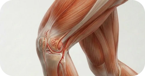
An ACL tear â a rupture of the anterior cruciate ligament inside the knee â is one of the most common and most recognizable athletic injuries in medicine. The pop, the immediate swelling, and the sense that the knee gave way are classic. If this happened to you today, this week, or this month, you are not alone â and the path forward is clear.
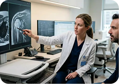
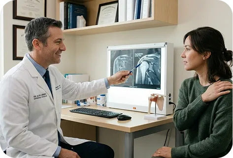
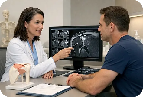
The MRI of a torn ACL tells a detailed story â not just that the ACL is torn, but how it tore, what else was injured at the same time, and what surgical planning is needed. Understanding your MRI findings helps you ask better questions at consultation and understand the surgical plan.
Â
ACL tear MRI is interpreted across six key dimensions:

A. Complete (Full-Thickness) ACL Tear â Grade 3
The ACL is completely disrupted â there is no continuous fiber bundle remaining between the femoral and tibial attachments. On MRI, the ligament appears absent, buckled, or as a wavy, discontinuous signal. A complete tear confirms ACL incompetence and is the primary surgical indication for reconstruction. Complete tears cannot spontaneously heal to a functional state.

A significant proportion of ACL fibers are torn, but some continuity remains. High-grade partial tears (> 50% of fibers torn) behave clinically like complete tears â they produce functional instability â and are typically managed as surgical candidates in active athletes. MRI characterization of partial tears can be challenging, and clinical examination (Lachman test laxity, pivot shift) is equally important in this determination.

A minority of fibers are torn. The ACL retains meaningful mechanical function. Low-grade partial tears may be managed conservatively in selected patients â particularly older, lower-demand adults â but active athletes in pivoting sports typically warrant surgical evaluation given the ongoing instability risk.

The tear occurs within the body of the ligament â between the femoral and tibial attachments. This is the most common ACL tear pattern and requires reconstruction (replacement with a graft). The torn ligament ends cannot be directly sutured back together â the tissue is not repairable in the traditional surgical sense.

The ACL tears at or very near its femoral attachment on the lateral wall of the intercondylar notch. Proximal tears â where the ligament end is in close proximity to its attachment and the tissue quality is good â are the primary indication for the BEAR (Bridge-Enhanced ACL Repair) procedure, which stimulates the ACL to heal in situ rather than replacing it with a graft. Proximal tears must be identified before surgery if BEAR is being considered.

The ACL tears at or near its tibial attachment. Less common than mid-substance or proximal tears. Reconstruction is typically performed.

A bony fragment from the tibial spine avulses with the ACL â the ligament itself remains intact but the bone attachment is disrupted. Bony avulsions â particularly in adolescents â may be amenable to direct fixation (screw or suture fixation of the fragment back to the tibia) rather than full reconstruction. This is an important distinction that guides surgical approach.
ACL tears are accompanied by meniscal tears in approximately 50â70% of cases. MRI characterizes both the ACL tear and the meniscal status, which significantly affects surgical planning â specifically whether meniscal repair or meniscectomy is performed concurrently with ACL reconstruction.
Â
Chronic ACL deficiency is particularly associated with progressive medial meniscal tearing as the secondary stabilizers are overloaded. A medial meniscal tear identified at the time of ACL reconstruction is repaired or partially removed at the same surgical procedure.
Acute ACL tears are frequently accompanied by lateral meniscal tears â particularly lateral meniscal root tears or radial tears â from the same high-energy event that caused the ACL rupture. Lateral meniscal preservation through repair is a high priority at ACL reconstruction.
A large, displaced bucket handle tear of the meniscus trapping between the femur and tibia â producing a locked knee â requires urgent surgical management alongside ACL reconstruction. A locked knee from a bucket handle tear is the only knee ligament injury scenario that represents a true surgical urgency.

High-energy ACL tears â from football tackles, skiing crashes, and contact sport injuries â frequently involve additional ligament damage that must be identified and addressed.
Â
The most common concurrent ligament injury with ACL tears. Grade I and II MCL injuries typically heal non-operatively while the ACL reconstruction is performed. Grade III MCL tears may require concurrent repair or reconstruction.
Bone bruises â impaction injuries to the subchondral bone present in the majority of acute ACL tears â confirm the high-energy pivot mechanism and matter for two reasons: they explain the acute pain and stiffness following injury, and they indicate that the overlying articular cartilage has been acutely loaded, which is why allowing the bone bruise to resolve over 2â6 weeks before ACL reconstruction is associated with better early outcomes than proceeding to immediate surgery.

MRI provides an initial assessment of articular cartilage across all three knee compartments, and full-thickness defects identified pre-operatively may require concurrent cartilage procedures â microfracture, chondroplasty, or osteochondral grafting â at the time of ACL reconstruction; this is also one of the primary reasons surgical reconstruction is generally preferred over prolonged non-operative management in active patients, as chronic ACL deficiency is strongly associated with progressive cartilage deterioration and secondary meniscal damage that adds complexity and risk to any eventual reconstruction.
MRI identifies the tear pattern, concurrent injuries, and the articular status of the knee. But the surgical plan â graft selection, concurrent procedures, approach to LET or BEAR â requires integration of the MRI with the patient’s age, sport, activity level, instability symptoms, and return-to-sport timeline. A fellowship-trained knee specialist combines all of these factors into the optimal ACL reconstruction plan for each individual patient.
*Same-day consultations may be available

Even though an MRI provides valuable structural information, it does not establish a complete diagnosis. A rotator cuff tear must be interpreted in the context of strength, movement quality, symptom behavior, and the physical demands placed on your shoulder â both now and over time.âĻ
Â
A shoulder specialist integrates these factors through comprehensive evaluation before defining the most appropriate path forward.

No â not functionally. A complete ACL tear will not functionally heal on its own â the ligament has extremely limited intrinsic healing capacity, and while scar tissue may form across the tear and provide some degree of stability, it is mechanically inferior to both the native ACL and a reconstructed graft; the practical question for a young active patient is therefore not whether the ACL heals (it won’t) but whether the secondary stabilizers can adequately compensate for its absence in the context of their specific sport and functional demands.

This is the most important argument for surgical reconstruction in active patients â and it is often underappreciated:
Â

Athletic and sport:
Â
Occupational and daily function:
Not every ACL tear requires immediate surgery â older, sedentary adults who avoid pivoting sports may achieve satisfactory function with bracing and rehabilitation â but for active adults, athletes, and anyone who needs to trust their knee under physical demand, conservative management has well-defined limits: physical therapy cannot reattach the ACL to its attachments, and while the dynamic stabilizers can be strengthened to partially compensate for its absence, that compensation has a ceiling that is typically not high enough for patients in pivoting sports or physically demanding occupations.
Â
Signals That Conservative Management Is Not Working:
Â
ACL tear management is individualized to tear pattern (complete/partial/proximal avulsion), concurrent injuries, patient age, sport and activity level, return-to-sport timeline, and prior surgical history. The full spectrum â from BEAR procedure for selected proximal tears to complex revision reconstruction with bone grafting â is available at The Joint Preservation Center.
Conservative management is appropriate in selected patients:
Â
Â
Conservative management requires a structured rehabilitation program â quadriceps strengthening, hamstring co-activation, proprioceptive training, and sport-specific neuromuscular training â to maximize the dynamic compensation for ACL absence. A functional knee brace may provide additional positional stability.
Arthroscopic ACL reconstruction with autograft â tissue taken from the patient’s own body â is the standard surgical treatment for complete ACL tears in active adults and athletes. The torn ACL is not repaired (sutured back together) â it is replaced with a graft that is placed through tunnels drilled in the femur and tibia, anchored with fixation hardware, and remodels over time into a functional ligament (ligamentization).
The central third of the patellar tendon â with a bone plug at each end from the patella and tibial tubercle â is harvested and used as the ACL graft. The bone-to-bone healing at the femoral and tibial tunnels is the fastest healing of any graft fixation, and BPTB grafts have a long track record of excellent outcomes in contact athletes.
Â
Considerations: Donor site morbidity includes anterior knee pain and kneeling discomfort at the patellar tendon harvest site in some patients. This is the most relevant limitation of patellar tendon grafts. For patients whose sport or occupation does not involve frequent kneeling, donor site discomfort is less clinically significant.
Â
Best suited for: Contact athletes (football, rugby, wrestling, MMA), patients with high physical demands, and patients for whom graft strength and bone-to-bone healing speed are the primary priorities.

The semitendinosus â and sometimes the gracilis â tendon from the medial aspect of the knee is harvested, folded multiple times, and used as a quadrupled or tripled graft bundle. Hamstring grafts avoid the patellar tendon donor site and produce comparable long-term outcomes to patellar tendon grafts in most patient populations.
Â
Considerations: Hamstring harvest may produce some weakness in knee flexion that is typically transient. Graft size is dependent on the diameter of the harvested hamstring tendons â smaller tendons produce smaller grafts with higher re-tear risk, which is a relevant consideration in younger patients and adolescents. Hamstring grafts attach through cortical buttons or interference screws and rely on soft tissue healing in bone tunnels.
Â
Best suited for: Recreational athletes, patients concerned about anterior knee pain, and patients where hamstring tendon size is adequate for the desired graft diameter.

The central portion of the quadriceps tendon â with or without a patellar bone plug â is harvested from the front of the thigh and used as the ACL graft. Quadriceps tendon grafts are among the largest autografts available and have gained significant traction in the ACL reconstruction literature over the past decade.
Â
Key advantages of quad tendon grafts: No anterior knee pain or kneeling discomfort (the harvest site is proximal to the kneecap rather than at the patellar tendon). Large graft diameter â important for reducing re-tear risk, particularly in younger patients and adolescents. The quad tendon harvest does not significantly weaken the quadriceps mechanism when appropriately performed.
Â
Best suited for: Younger athletes, adolescents, patients concerned about patellar tendon donor site morbidity, and revision surgery where prior patellar tendon or hamstring graft has been used.

Allograft ACL reconstruction uses cadaveric donor tissue, eliminating donor site morbidity entirely and allowing earlier post-operative comfort â but carries a significant limitation: in patients under 25â30, allograft re-tear rates are substantially higher than autograft in high-demand athletes, as processed and sterilized tissue remodels more slowly and incompletely in younger patients, with published studies consistently showing higher failure rates in young, active, pivoting sport athletes; allograft is therefore most appropriate for recreational athletes over 35â40 with moderate return-to-sport demands, revision cases where prior autografts have been exhausted, or specific circumstances where autograft harvest is not feasible â and is not generally recommended as a primary graft choice for competitive athletes under 25.
The BEAR (Bridge-Enhanced ACL Repair) procedure is a relatively novel surgical approach that â in selected patients â allows the ACL to heal in its native position rather than being replaced with a graft. The BEAR procedure uses a bioactive scaffold, sutured into the torn ACL ends and saturated with the patient’s own blood, to bridge the gap between the torn ends and facilitate biological healing.
BEAR is appropriate only for a specific subset of ACL tears:
Â
BEAR is NOT appropriate for mid-substance tears (the most common pattern), distal tears, chronic tears, or patients without an adequate ACL stump. Patient selection requires careful MRI evaluation and clinical assessment.
Â
The current BEAR evidence base shows promising short and medium-term outcomes comparable to hamstring graft reconstruction in appropriately selected patients. Long-term outcomes data continues to accumulate, and BEAR remains a specialized procedure that is not universally available or indicated. We evaluate every ACL tear patient for BEAR candidacy when appropriate.

Lateral extra-articular tenodesis (LET) is an adjunct procedure performed concurrently with ACL reconstruction â not as a standalone treatment. LET uses a strip of the iliotibial band (IT band) to create an additional restraint on the lateral side of the knee, augmenting rotational stability beyond what the ACL graft alone provides.
Published evidence â particularly the STABILITY trial â shows that ACL reconstruction with LET significantly reduces graft re-tear rates compared to ACL reconstruction alone in high-risk patients. LET is indicated when:
Â
Â
LET adds modest surgical time and a small additional lateral incision to the ACL reconstruction procedure. It does not meaningfully prolong overall recovery or return to sport timelines. For appropriately selected patients, the evidence for LET as a re-tear risk reduction strategy is compelling.

One of the most distinctive features of ACL surgery â compared to almost every other orthopedic procedure â is that patients actively research graft selection before their first surgical consultation. No other procedure in this series generates the volume of graft-comparison searches that ACL does. Understanding how graft selection decisions are made helps patients engage productively at consultation.
ACL tears are increasingly common in adolescent athletes â particularly in pivoting sports such as soccer, basketball, football, volleyball, and gymnastics, with female athletes at significantly higher risk than males due to anatomic, hormonal, and neuromuscular factors â and their surgical management in skeletally immature patients requires specialized physeal-sparing techniques, as standard adult ACL reconstruction that drills tunnels directly through the open growth plates can cause growth disturbance, limb length discrepancy, or angular deformity, which is not a theoretical concern but a documented complication of applying adult techniques to skeletally immature patients.
Physeal-sparing ACL reconstruction modifies the tunnel placement and graft passage to avoid crossing the open growth plates of the distal femur and proximal tibia â the most active growth regions in the knee. Physeal-sparing techniques maintain ACL stability while minimizing growth plate exposure.
Â
The appropriate technique depends on the patient’s skeletal maturity â assessed by bone age X-ray and Tanner staging â and the specific anatomy of the growth plates:
Â

The critical question in adolescent ACL management â whether to operate promptly or defer until skeletal maturity â is increasingly answered in favor of earlier intervention, as the risk of secondary meniscal and cartilage damage from ongoing instability in an active adolescent is substantial and deferral, while avoiding growth plate risk, often results in significant secondary joint damage during the waiting period; for skeletally immature patients active in pivoting sports, physeal-sparing ACL reconstruction is strongly preferred over prolonged non-operative management, while for those approaching skeletal maturity the growth plate risk is lower and standard techniques become increasingly appropriate â making evaluation by a surgeon experienced in both pediatric growth plate anatomy and physeal-sparing techniques essential.
ACL graft failure â the graft rupturing after a technically successful initial reconstruction â is a recognized complication. Published graft re-tear rates vary by graft type, patient age, and sport: in competitive young athletes, re-tear rates can reach 15â25%, particularly with allograft use in this population. Understanding why the graft failed is the essential first step before revision surgery.
Revision ACL surgery is more complex than primary reconstruction:
Â
Â
A second opinion before revision ACL surgery is always appropriate. Understanding why the first surgery failed â which requires review of the original operative report, current MRI, and CT tunnel measurement â is essential before committing to a revision approach.
High-energy ACL tears â from football tackles, skiing falls, soccer pivots, and direct contact â frequently involve additional structural damage that must be identified and addressed at surgery. Addressing all pathology in a single procedure minimizes anesthesia exposure and allows coordinated rehabilitation.
Meniscal tears accompany ACL tears in approximately 50â70% of cases. Meniscal preservation through repair is the highest priority when the tear pattern and tissue quality allow â the long-term consequences of meniscus loss are significant, and every effort is made to repair rather than remove meniscal tissue.
Â

The terrible triad is the classic combined knee injury from contact sport: ACL, medial collateral ligament (MCL), and medial meniscal tear. Management is individualized â MCL tears are typically managed non-operatively (bracing) while the ACL and meniscus are addressed surgically. Grade III MCL tears with persistent laxity may require surgical repair or reconstruction.

Combined ACL and PCL tears produce severe multiligament instability that requires comprehensive surgical planning. These are among the most complex knee reconstructions performed â typically requiring staged or combined reconstruction of both cruciate ligaments and any concurrent collateral ligament injuries.

Articular cartilage defects identified at ACL reconstruction are addressed concurrently when appropriate â chondroplasty for shallow defects, microfracture or osteochondral grafting for full-thickness defects. Cartilage procedures may affect rehabilitation timing and return-to-sport milestones.

ACL reconstruction recovery is one of the most extensively studied rehabilitation processes in sports medicine, governed by objective milestones rather than calendar dates â and understanding its biological foundation helps athletes commit to the full process: the transplanted graft must undergo ligamentization, a remodeling process in which graft tissue is progressively replaced with ACL-like collagen over approximately 12â24 months, leaving the graft at its weakest during the 3â6 month window; this biological reality â not surgeon conservatism â is why return to pivoting sport before 9â12 months is associated with higher re-tear rates.
Phase 1: Immediate Post-Op (Weeks 0â2)
Crutches for weight bearing protection. Ice and elevation for swelling control. Range-of-motion exercises begin immediately to prevent arthrofibrosis (scar-induced stiffness). Early quad activation is essential â the quad must begin firing immediately.
Â
Phase 2: Early Rehabilitation (Weeks 2â6)
Progressive weight bearing â most patients achieve full weight bearing by 4â6 weeks. Crutches typically discontinued. Range of motion restoration continues. Quad and hamstring strengthening progresses.
Â
Phase 3: Neuromuscular and Strengthening (Weeks 6â16)
Progressive strengthening of quads, hamstrings, hip, and core. Proprioceptive and neuromuscular training. Sport-specific movement patterns begin without competition load.
Â
Phase 4: Sport-Specific Training (Months 4â9)
Running program progresses. Sport-specific drills â cutting, pivoting, deceleration â begin progressively. Return-to-sport testing criteria are assessed.
Â
Phase 5: Return to Sport (Month 9â12+)
Return to full sport is cleared based on objective criteria: limb symmetry index (LSI) on strength testing above 90%, hop testing symmetry, and psychological readiness. Return to contact and pivoting sport should not occur before 9 months at the earliest, with many surgeons and sports medicine physicians recommending 12 months for the highest-risk patients.
Published data consistently show that athletes who return to sport before meeting strength and functional testing milestones have significantly higher re-tear rates than those who meet objective criteria before return. Calendar-based return decisions â ‘it’s been 9 months, so I’m cleared’ â are inferior to milestone-based decisions that require the knee to actually prove it is ready.
Â
The primary return-to-sport testing battery includes: isokinetic quad and hamstring strength testing (limb symmetry index > 90%), hop tests (single-leg hop, triple hop, crossover hop â symmetry > 90%), and dynamic stability assessments. Psychological readiness â the patient’s confidence in the knee during sport â is also assessed and is an independent predictor of re-tear risk.

Modern ACL reconstruction techniques are associated with:
High rates of return to sport and pre-injury activity
Restored knee stability and rotational control
Improved functional outcome scores
High patient satisfaction
1
We evaluate your ACL tear pattern, concurrent injuries, knee laxity, sport and activity level, age, and return-to-sport goals. For acute injuries, we provide same-day or same-week evaluation. For patients researching graft selection, we review MRI, explain each graft option in the context of your specific anatomy and sport, and make a clear recommendation.
2
We recommend the most appropriate reconstruction approach â patellar tendon, hamstring, quad tendon, BEAR (if eligible), allograft (if indicated), with or without LET â based on your age, sport, anatomy, and prior surgical history. We discuss the evidence for each option transparently.
3
If surgery is recommended, our fellowship-trained knee surgeons perform the appropriate reconstruction â primary ACL reconstruction with your selected graft, physeal-sparing technique for skeletally immature patients, LET when indicated, concurrent meniscus and cartilage procedures as needed, and revision reconstruction for failed prior surgery.
4
Our surgeons work closely with physical therapists to guide a phased, milestone-based rehabilitation program â from early quad activation and range of motion through sport-specific training and objective return-to-sport testing.
5
Return to full sport is cleared based on objective criteria â strength symmetry, hop test performance, and psychological readiness â not calendar dates alone. The goal is a durable return to competition with the lowest possible risk of re-tear.
This long-term follow-up helps us understand how knees perform beyond the first year â including graft re-tear rates, return-to-sport rates and levels, limb symmetry recovery, contralateral ACL tear rates, and long-term joint health. These insights allow our surgeons to continually refine patient selection, graft choice, LET indications, and rehabilitation protocols to support the most durable outcomes over time â particularly for young athletes whose knee health must last decades.

1
Elite surgeons with decades of experience, incentivized to do the right thing
2
Prevent future surgeries
3
Heal with advanced, minimally invasive techniques
4
Preserve your natural joints, whenever possible
5
Seamless coordination from injury to recovery
6
Premium personalized care, made accessible
7
All patient outcomes tracked for 5 years
For active adults and athletes who participate in pivoting, cutting, or contact sports â yes, ACL reconstruction is generally recommended. The ACL does not heal functionally after a complete tear, and the dynamic compensators (hamstrings, quad) cannot indefinitely prevent the pivot-shift instability that a torn ACL creates.
Â
For older, less active adults who do not participate in pivoting sports, conservative management with bracing and rehabilitation may be a reasonable long-term option. The critical consideration in conservative management is the risk of secondary meniscal and cartilage damage from ongoing instability â which can make eventual surgery more complex.
The ‘best’ graft depends on your age, sport, anatomy, and prior surgical history. General guidance:
Â
Graft selection is discussed in detail at consultation based on your specific MRI, anatomy, and goals. See Section 9 for the complete decision framework.
The BEAR (Bridge-Enhanced ACL Repair) procedure is a specialized approach that allows the ACL to heal in its native position â using a bioactive scaffold saturated with the patient’s own blood to bridge the torn ends â rather than replacing the ligament with a graft. BEAR is only appropriate for specific patients: those with proximal (femoral-side) tears, acute injuries, and an adequate ACL stump remaining. It is not appropriate for mid-substance tears or chronic tears. We evaluate every patient for BEAR candidacy when the tear pattern suggests eligibility.
Lateral extra-articular tenodesis (LET) is an adjunct procedure performed alongside ACL reconstruction that adds rotational stability using a strip of the IT band on the outer knee. LET significantly reduces graft re-tear rates in high-risk patients: young athletes under 25, contact sport players, patients with a positive pivot shift examination, and revision reconstruction cases. LET adds minimal surgical time and does not meaningfully prolong recovery. If you fall into a high-risk category, LET is a meaningful re-tear risk reduction strategy.
Yes â and in most active adolescent athletes with a complete ACL tear and ongoing instability, surgery is the appropriate recommendation. The concern about ACL surgery in teenagers is growth plate safety, which is addressed through physeal-sparing reconstruction techniques that avoid drilling through the open growth plates.
Â
Physeal-sparing ACL reconstruction has an established safety and efficacy record. The risk of waiting â allowing an active adolescent to sustain progressive meniscal and cartilage damage from repeated giving-way episodes â typically outweighs the carefully managed growth plate risk of expert physeal-sparing surgery. Seek evaluation from a surgeon with specific experience in adolescent ACL reconstruction and physeal-sparing techniques.
Graft failure requires evaluation of why the graft failed before planning revision surgery. A current MRI and CT tunnel measurement are essential. Options include:
Â
A second opinion before revision ACL surgery is strongly encouraged. Understanding the failure mechanism is essential to avoiding the same outcome with the revision.
Most athletes return to full contact and pivoting sport at 9â12 months after ACL reconstruction. The timeline is governed by objective milestones â quad and hamstring strength symmetry above 90%, hop test symmetry â not by calendar dates alone. Return before 9 months is associated with higher re-tear rates. Many sports medicine specialists now recommend 12 months for contact athletes and young patients.
Absolutely â and for graft selection, BEAR candidacy, LET indication, or revision surgery planning, a second opinion from a fellowship-trained knee surgeon experienced in these decisions is valuable. We offer second opinion consultations, including virtual consultations for patients who cannot travel.
The Joint Preservation Center accepts most PPO insurance plans that have out-of-network benefits:
If you have a PPO insurance plan with out-of-network benefits:
Note: If you have Medicare, Medicaid, TRICARE, or VA programs, or if your PPO does not have out-of-network benefits, you can still see our specialists and the surgery center will still be in-network. In this case, our specialists charge $250 for the initial office visit (all follow ups are included). Surgery is typically in the range of $6K â $8K depending on what you need done.
If you are unsure whether your plan is accepted, our team can verify your coverage before your appointment.
*Same-day consultations may be available
*Online visits available