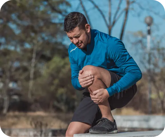
When knee pain or swelling doesn’t go away, especially after an injury, it can be frustrating — and even concerning. One possible cause is something called an Osteochondral lesion (also known as an Osteochondritis Dissecans, or OCD lesion).
Although the name sounds complicated, understanding this condition and its treatment options can help you feel more confident and in control of your recovery.
General Points
What Is an Osteochondral/OCD Lesion?
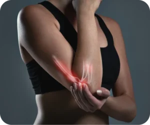
An osteochondral lesion occurs when a small area of bone and its overlying cartilage inside a joint — most often the knee, but sometimes the ankle or elbow — becomes damaged.
This damage may happen after an injury, repetitive stress, or from reduced blood flow to the bone beneath the cartilage. Over time, this weak spot can cause pain, swelling, and even a small fragment of cartilage and bone to loosen inside the joint.
In younger, active adults under 65, it’s one of the most common causes of persistent joint pain, particularly after sports injuries or repeated impact activities.
Early diagnosis and treatment are crucial because cartilage doesn’t heal on its own — and when left untreated, the condition can worsen, increasing the risk of early arthritis.
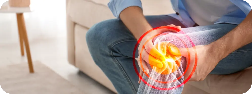
Common Symptoms
Patients with osteochondral or OCD lesions usually report symptoms that can vary depending on the size and location of the lesion, but the most frequent include:
- Deep, aching joint pain, often worsened by activity
- Swelling or stiffness, especially after exercise
- Locking or catching sensations in the joint
- Feeling of instability or “giving way” during movement
- Decreased range of motion or discomfort with weight-bearing
Many patients describe the pain as intermittent — good days and bad days — which often leads them to delay seeking care. However, early evaluation by an orthopedic specialist can make a big difference in outcomes.
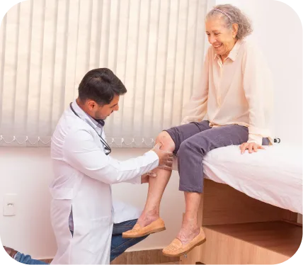
How Is an Osteochondral/OCD Lesion Diagnosed?
Diagnosis typically starts with a thorough physical examination. Your orthopedic surgeon will assess swelling, tenderness, range of motion, and joint stability.
Imaging Studies
To confirm the diagnosis and determine the extent of damage, several imaging tools are used:
- X-rays – to visualize bone alignment and rule out fractures
- MRI scans – to see the cartilage and bone in detail, and to evaluate if the fragment is still attached or loose
- CT scans – sometimes used to assess bone healing or plan surgery
These tests help the surgeon determine whether the lesion is stable (attached) or unstable (loose or displaced) — a key factor in deciding whether surgery is necessary.
Treatment Options for Osteochondral/OCD Lesions
Treatment depends on several factors — including lesion size, stability, age, and activity level.
Non-Surgical Management
For small, stable lesions, especially in younger patients, non-surgical care may be tried first. This may include:
- Activity modification (avoiding high-impact sports)
- Bracing or limited weight-bearing
- Physical therapy to restore strength and mobility
- Anti-inflammatory medications
However, if symptoms persist or imaging shows an unstable fragment or larger cartilage defect, surgical intervention is often the best solution to restore joint health and prevent further degeneration.
Surgical Treatment Options
Arthroscopic vs. Open Osteochondral Pinning vs. Cartiheal or MACI
When surgery is needed, the goal is to restore the damaged cartilage and underlying bone, allowing for smooth joint motion and long-term function. Depending on the case, your surgeon may recommend one of the following procedures.
Arthroscopic Osteochondral Pinning
What it is
Arthroscopic pinning is a minimally invasive procedure performed through small incisions using a camera (arthroscope). The surgeon identifies the loose or unstable cartilage fragment and reattaches it to the bone using small, bioabsorbable pins or screws.
When it’s recommended
This approach is typically used when the osteochondral fragment is still viable (alive) and large enough to be fixed back into place, especially in younger or active adults.
Benefits
- Minimally invasive — less pain and quicker healing
- Preserves the patient’s natural cartilage
- Small incisions and minimal scarring
- High rates of successful healing when done early
Success rates
Studies show 80–90% success in fragment healing and pain relief, especially when performed before the cartilage deteriorates significantly.
Open Osteochondral Pinning (or Fixation)
What it is
In some cases, particularly when the lesion is large, deep, or difficult to access arthroscopically, an open approach is preferred. The surgeon makes a slightly larger incision to directly visualize the lesion, realign the bone and cartilage, and secure it using specialized bioabsorbable pins or small screws.
When it’s recommended
- Large or complex lesions
- Detached fragments that need precise repositioning
- Failed prior arthroscopic fixation
Benefits
- Direct visualization allows more accurate placement of the fragment
- Reliable fixation for larger or irregular defects
- Restores the native cartilage surface and joint function
Success rates
Most studies report 85–95% good to excellent outcomes, particularly when combined with proper rehabilitation and activity modification during recovery.
Cartiheal (Agili-C) or MACI (Matrix-Induced Autologous Chondrocyte Implantation)
For larger or more chronic defects where the cartilage is no longer viable, regenerative cartilage restoration procedures are an advanced solution.
Cartiheal (Agili-C):
Cartiheal uses a biological scaffold made of natural minerals that supports new bone and cartilage growth. It doesn’t require cell harvesting and can be used in both osteochondral and purely cartilage lesions.
Benefits
- Single-stage surgery
- Promotes true cartilage regeneration (not scar tissue)
- Suitable for a wide range of lesion sizes
- Excellent outcomes for pain relief and mobility
Success rates
Early studies show over 90% patient satisfaction and sustained improvement for up to 5 years.
MACI (Matrix-Induced Autologous Chondrocyte Implantation):
MACI is a two-step regenerative procedure. In the first step, a small sample of your healthy cartilage cells is taken and grown in a lab. In the second step, these cells are implanted on a collagen membrane and placed over the damaged area to regrow cartilage naturally.
Benefits
- Uses your own cells for long-term cartilage restoration
- Restores smooth, functional cartilage surface
- Especially useful for large or chronic defects
Success rates
Clinical trials report 80–90% good to excellent results, with long-term durability for 10+ years when proper rehabilitation is followed.
Long-Term Outlook
With modern surgical techniques such as Arthroscopic and Open Osteochondral Pinning, Cartiheal, and MACI, most patients experience significant improvement in joint function, reduced pain, and a return to normal activity — even competitive sports. The key to success is early diagnosis, appropriate surgical planning, and a guided rehabilitation program.
When to See an Orthopedic Specialist
If you’ve been diagnosed with an Osteochondral/OCD lesion or continue to experience knee pain that doesn’t improve with rest, consulting an experienced orthopedic surgeon is the best next step.
Prompt evaluation can prevent worsening damage and help you explore the most effective treatment — from minimally invasive pinning to advanced cartilage restoration procedures.
Take the First Step Toward Full Recovery
Living with an Osteochondral lesion doesn’t mean giving up the activities you love. With modern techniques like Arthroscopic vs. Open Osteochondral Pinning vs. Cartiheal or MACI, patients today have multiple effective options to restore their joint health and return to an active lifestyle — safely and confidently.
If you’re struggling with persistent knee pain or have recently been diagnosed, schedule a consultation with an orthopedic specialist to discuss your personalized treatment plan and take the first step toward lasting recovery.