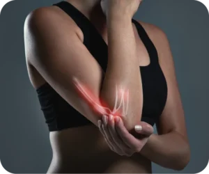
Chondral defects, or injuries to the cartilage that covers the joints, are common yet often overlooked conditions that can lead to serious long-term consequences if not addressed early. Cartilage plays a critical role in allowing smooth and painless joint movement.
When this cartilage becomes damaged—whether due to trauma, wear and tear, or degeneration—it can significantly affect your mobility and quality of life. In this article, we’ll discuss what a chondral defect/injury is, how it’s diagnosed, and the most effective treatments available, including surgical options like Arthroscopic, Open Cartilage Restoration (Cartiheal), and Implantation (MACI).
General Points
What Is a Chondral Defect/Injury?
A chondral defect is a type of cartilage injury that affects the smooth, elastic tissue lining your joints, especially the knee, hip, and ankle. This cartilage, known as articular cartilage, is vital for reducing friction between bones and absorbing shock during movement. When it becomes damaged, the bones in the joint rub against each other, causing pain, stiffness, and potentially leading to further joint deterioration.
Chondral defects can be caused by a variety of factors, including:
- Trauma or injury: A direct blow or sports-related injury, like twisting the knee, can cause cartilage damage.
- Wear and tear (Osteoarthritis): Age and repetitive movements can gradually wear down cartilage, making it more susceptible to injury.
- Overuse: Frequent high-impact activities or repetitive stress can lead to cartilage degradation over time.
If left untreated, a chondral defect can evolve into more severe conditions, such as osteoarthritis, leading to chronic pain, limited range of motion, and even permanent joint damage.
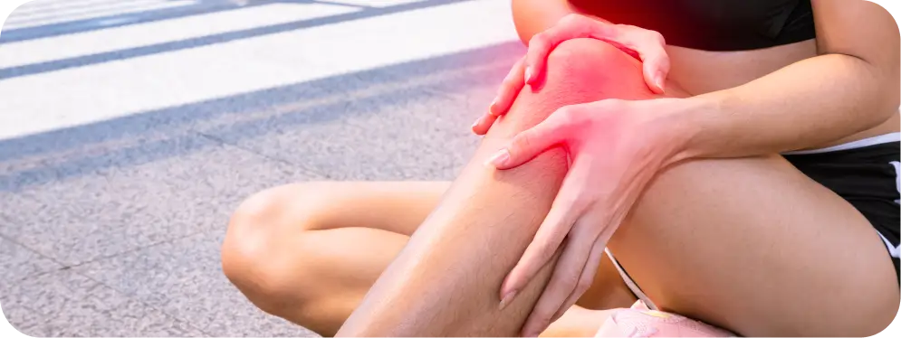
Symptoms of Chondral Defect/Injury
The symptoms of a chondral defect can vary depending on the location and severity of the injury. However, the most common signs include:
- Pain: You might feel pain around the affected joint, especially when performing certain movements or after physical activity.
- Swelling: The injured area may swell up, which is a result of inflammation in response to the damage.
- Stiffness: You may experience a feeling of tightness or decreased range of motion in the joint, particularly after periods of rest.
- Locking or catching: In some cases, damaged cartilage can cause the joint to “catch” or “lock,” making it difficult to move the joint properly.
- Instability: The joint might feel unstable or weak, as if it could “give way” under pressure.
If you notice these symptoms, it’s important to seek medical attention early to prevent the condition from worsening and to explore treatment options that can relieve pain and restore function.
How Is Chondral Defect/Injury Diagnosed?
Diagnosing a chondral defect involves a thorough examination by an orthopedic specialist. Here’s what to expect during the diagnostic process:
Physical Exam
Your doctor will first perform a physical exam to assess the affected joint’s range of motion, tenderness, and any signs of instability. They may ask you about any recent injuries, physical activities, or family history of joint issues, such as osteoarthritis.
Imaging Tests
To confirm the diagnosis, your doctor will likely order imaging tests, including:
- X-rays: While X-rays can’t directly show cartilage damage, they help rule out other causes of pain, such as fractures or joint misalignment.
- MRI (Magnetic Resonance Imaging): This is the most accurate imaging technique for assessing cartilage damage. An MRI creates detailed images of both soft tissues and bone, allowing your doctor to see the extent of the chondral defect.
- Arthroscopy: In some cases, a minimally invasive procedure called arthroscopy might be performed to visualize the damage directly inside the joint. This involves inserting a small camera (arthroscope) through a tiny incision.
Once the extent of the cartilage damage is confirmed, your doctor will discuss treatment options based on the severity of the injury and your personal activity level.
Treatment Options for Chondral Defect/Injury
The treatment for a chondral defect will depend on several factors, including the size and location of the injury, your age, activity level, and overall health. There are both non-surgical and surgical treatment options available.
Non-Surgical Treatments
For minor chondral defects, non-surgical treatments may be recommended to reduce symptoms and promote healing. These can include:
- Rest and Activity Modification: Limiting high-impact activities to avoid further stress on the joint.
- Physical Therapy: Targeted exercises to strengthen the muscles around the joint, improve stability, and reduce strain on the cartilage.
- Medications: Nonsteroidal anti-inflammatory drugs (NSAIDs) can help manage pain and inflammation.
- Injections: Corticosteroid injections or hyaluronic acid injections may be used to reduce inflammation and improve joint lubrication.
If non-surgical treatments don’t provide sufficient relief, surgical options might be considered. Below, we will focus on three primary surgical procedures: Arthroscopic Surgery, Open Cartilage Restoration (Cartiheal), and Implantation (MACI).
Surgical Treatment Options
When non-surgical treatments aren’t enough to resolve a chondral defect, surgical intervention becomes necessary. Depending on the severity of the cartilage damage, different procedures may be recommended. Below, we will discuss three common surgical treatments for chondral defects: Arthroscopic Surgery, Open Cartilage Restoration (Cartiheal), and Implantation (MACI).
Arthroscopic Surgery
How It Works
Arthroscopic surgery is a minimally invasive procedure that involves small incisions and the use of a tiny camera (arthroscope) to view and treat the cartilage damage. This approach is ideal for smaller defects or when the damage is located in areas that are easy to access.
- Cartilage Debridement and Microfracture: During the procedure, the surgeon may perform “debridement,” which involves cleaning the damaged area by removing any loose cartilage or debris. In cases where the damage is deeper, the surgeon may use a technique called microfracture, where tiny holes are created in the bone to stimulate the growth of new cartilage.
- Shaping and Smoothing: Sometimes, the surgeon will also smooth or reshape the cartilage to ensure that the joint moves as fluidly as possible. This can improve function and reduce pain.
When It’s Recommended
Arthroscopic surgery is typically recommended for patients with smaller cartilage defects, especially when the damage is confined to one specific area. It is also the go-to treatment for individuals with minimal joint degeneration who want to preserve as much natural cartilage as possible.
Success Rates
Arthroscopic surgery has a high success rate for small defects, with studies showing that most patients experience significant pain relief and functional improvement. The minimally invasive nature of the procedure, combined with short recovery times, makes it a popular option for many patients. However, larger defects or deep cartilage injuries may not be fully addressed with arthroscopy alone, which is why additional treatments might be considered.
Open Cartilage Restoration (Cartiheal)
How It Works
Cartiheal is a relatively new and innovative surgical approach designed to treat larger or deeper cartilage defects. It involves the implantation of a bioactive collagen scaffold, which serves as a foundation for new cartilage growth. This technique encourages the body’s natural healing process and allows the joint to regenerate its own cartilage over time.
- Bioactive Collagen Scaffold: The procedure involves placing a collagen scaffold into the damaged area of the cartilage. This scaffold is impregnated with growth factors that stimulate the body’s ability to heal itself by promoting the regeneration of cartilage cells.
- Bone and Cartilage Integration: The scaffold integrates with the surrounding bone and cartilage to form a stable foundation, encouraging natural healing and minimizing the risk of future damage. Over time, the body gradually replaces the scaffold with its own new cartilage.
When It’s Recommended
Cartiheal is often recommended for patients with larger chondral defects or those whose injuries are too extensive for traditional arthroscopic surgery. It’s also a great option for individuals who have previously undergone arthroscopic surgery but are still dealing with persistent cartilage damage.
Success Rates
The Cartiheal procedure has shown promising results, particularly for patients with medium-to-large defects. Studies have reported excellent cartilage regeneration and improvements in joint function and pain reduction. The success of this procedure is largely due to its ability to regenerate cartilage using the body’s own cells and natural healing mechanisms.
Implantation (MACI)
How It Works
Matrix-induced Autologous Chondrocyte Implantation (MACI) is an advanced technique for repairing larger cartilage defects, and it involves using your own cartilage cells to create new cartilage. Here’s how it works:
- Harvesting Cartilage Cells: The first step in the MACI procedure involves collecting healthy cartilage cells from an area of your joint that is less weight-bearing. These cells are sent to a lab, where they are cultured and multiplied to create a larger supply.
- Cell Implantation: Once the cells are ready, they are implanted onto a biodegradable scaffold and inserted into the damaged cartilage area. The scaffold provides support for the new cells and helps them integrate into the joint over time.
When It’s Recommended
MACI is most beneficial for individuals with significant cartilage damage in weight-bearing joints, such as the knee, where smaller surgical options may not be effective. It is typically recommended for patients who have not responded to other treatments, including arthroscopic surgery or microfracture.
Success Rates
MACI offers excellent long-term outcomes, particularly for those with large defects or significant cartilage loss. Studies have shown high success rates, with most patients experiencing substantial improvements in joint function, pain relief, and a significant reduction in the need for further surgeries. The procedure’s success is largely due to its ability to create biologically integrated, durable cartilage that mimics natural tissue.
What to Expect After Surgery
The recovery process for cartilage restoration surgery varies depending on the procedure, the extent of cartilage damage, and the patient’s individual health. Here’s what you can generally expect after each type of surgery:
Arthroscopic Surgery
With arthroscopic surgery, many patients experience significant relief from pain, improved joint function, and enhanced mobility. However, the results can vary depending on the size and location of the defect.
Open Cartilage Restoration (Cartiheal)
Cartiheal can be highly effective for larger defects, with many patients reporting excellent pain reduction and functional recovery in the long term.
Implantation (MACI)
MACI is a great option for large cartilage defects, and patients often report substantial improvements in joint function and pain relief. As the body gradually integrates the new cartilage, long-term benefits continue to improve.
By taking a comprehensive approach to both the surgery and recovery phases, patients can achieve the best possible outcome. Each treatment offers distinct advantages based on the severity of the injury, and understanding what to expect at each stage of recovery is crucial for long-term success.
Why Choose Expert Care
Chondral defects can be challenging, but with the right treatment, many patients experience a significant improvement in their joint function and quality of life. Whether you’re exploring non-surgical treatments or considering surgical options like Arthroscopic Surgery, Cartiheal, or MACI, it’s essential to consult with an experienced orthopedic specialist who can guide you through the most appropriate course of action.

Taking the first step toward treatment now can prevent further joint damage and improve your overall health. If you’ve been diagnosed with a chondral defect, don’t wait—schedule a consultation today to learn how we can help restore your joint function and relieve your pain.